Adipocytokine gene expression analysis in adipose tissue in patients with metabolic syndrome
Adipocytes, immune cells, and cells of the stromal vascular fraction are sources of many biologically active substances. The effects of most adipocytokines are well studied; however, the role of individual molecules secreted by adipose tissue in the pathogenesis of inflammation associated with metabolic syndrome (MS) and related diseases has not been fully elucidated and remains a matter of debate.
Aim — To evaluate the expression of adipocytokine genes in biopsies (fragments) of visceral adipose tissue of the gastrocolic omentum in patients with MS.
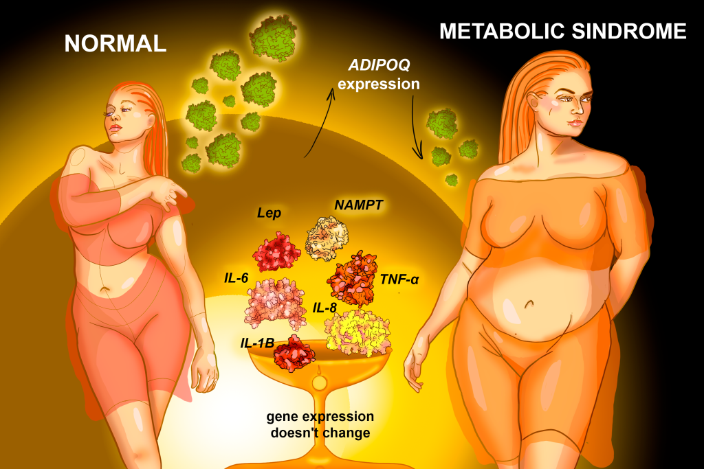
Materials and methods. The study included 19 patients: the main group — 14 patients with MS, the comparison group — 5 patients without MS. The level of expression of cytokine genes (IL1B, IL6, IL8, TNF-α) and peptide hormones (LEP (leptin), NAMPT (visfatin), RETN (resistin), ADIPOQ (adiponectin)) was studied in biopsies of adipose tissue of the greater omentum obtained during planned cholecystectomy for medical reasons by the level of matrix RNA using quantitative real-time PCR with preliminary development of complementary DNA in the reverse transcription reaction.
Results. There were no significant differences in the level of mRNA of IL-1B, -6, -8, TNF-α, as well as the hormones LEP, NAMPT, RETN in adipose tissue in patients with MS and in the comparison group (р > 0.05). Statistically significant differences were established only in the level of ADIPOQ gene expression: the level of ADIPOQ gene expression in adipose tissue was on average 2.8 times lower than in the comparison group (2.34 and 0.81 units, respectively, p < 0.05).
Conclusion. Analysis of the expression profiles of genes encoding key adipocytokines and of the functional and metabolic characteristics of visceral adipose tissue appears promising for identifying diagnostic and prognostic markers of MS and associated diseases.