
The effect of naturally occurring chelidonic acid on the morphofunctional status of internal organs: an experimental study
- DOI
- 10.5922/ATB-2026-2-1-7
- Pages
- 101-123
- To cite
- Gorokhova A. V., Nasibov T. F., Ryzhkova A. Yu., Leshenkova A. V., Porokhova E. D., Dzyuman A. N., Avdeeva E. Yu., Belousov M. V., Khlusov I. A. The effect of naturally occurring chelidonic acid on the morphofunctional status of internal organs: an experimental study. Advanced targets in Biomedicine. 2026;2(1):101—123. https://doi.org/10.5922/ATB-2026-2-1-7
Abstract
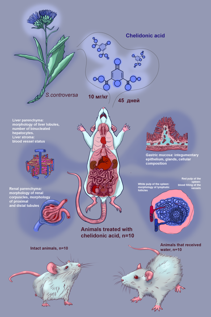
For the first time, a comprehensive in vivo evaluation of the effects of long-term (45 days) daily intragastric administration of water solution of naturally occurring small molecules of chelidonic acid (ChA) to Balb/c mice was conducted. The study was focused on the morphofunctional state of several key organs involved in xenobiotic metabolism and excretion, including the stomach, liver, kidneys, as well as an immunocompetent organ (spleen). The obtained results demonstrate the absence of pathological changes in these organs when the substance was administered orally at a concentration of 10 mg/ kg in 0.2 ml of solvent (distilled water) for 45 days. Moreover, as previously established, ChA and its calcium salt at the indicated dose exhibit specific (osteogenic) activity against mesenchymal stromal/stem cells both in vitro and in vivo. These findings indicate an absence of general toxicity to studied organs of a low concentration of ChA in lineage mice, suggesting that it may be a promising candidate for future clinical use. This opens up promising opportunities for further research and development of an active pharmaceutical substance that is entirely domestic in origin. This is of great importance for the advancement of the field of regenerative biomedicine, primarily in the area of bone tissue engineering.
Full text
Введение
Малые молекулы (ММ) — это органические соединения с низкой молекулярной массой, меньше 1000 Да, к которым относятся некоторые метаболиты, лекарственные препараты, моносахариды, вторичные мессенджеры и ксенобиотики [1]. Их применение в биомедицине и фармакологии обусловлено рядом преимуществ, определяющих перспективность использования: (1) благодаря низкой молекулярной массе ММ эффективно преодолевают клеточные мембраны, что обеспечивает их взаимодействие не только с внеклеточными, но и с внутриклеточными мишенями, включая регуляцию сигнальных путей [2—5]; (2) высокая диффузионная способность ММ способствует их накоплению в тканях с ограниченной васкуляризацией, что расширяет терапевтические возможности [4; 5]; (3) большинство ММ не требует сложных схем приема, а главный способ применения — пероральный [4; 5]; (4) быстрая абсорбция и предсказуемая фармакокинетика позволяют легче дозировать содержащие ММ препараты и гибко корректировать режимы терапии [4; 5]. Кроме того, синтез ММ в лабораторных условиях или их выделение из природных источников требуют меньших затрат по сравнению с макромолекулярными соединениями [6; 7] а их стабильность при хранении и транспортировке упрощает логистику [8; 9], делая ММ доступными даже для небольших исследовательских центров.
Хелидоновая кислота (ChA, молекулярная масса 184,1) — одна из ММ, весьма перспективная для биомедицинских приложений вследствие широкого спектра фармакологической активности [10]. Экспериментальные данные свидетельствуют о высокой иммуномодулирующей активности ChA на моделях воспалительных заболеваний, таких как язвенный колит у мышей, где она улучшала состояние животных, снижая уровни провоспалительных цитокинов IL- 6, TNF-α, а также экспрессию COX-2 и HIF-1α [11]. Ее противовоспалительные свойства также продемонстрированы в культуре человеческих кератиноцитов, где ChA нормализовала концентрации IL-1β, IL-6, IL-8 и TNF-α [12]. Кроме того, ChA показала противоаллергическое действие, снижая реакцию на аллергены, количество эозинофилов и уровни IL-1β и COX-2 у мышей с ринитом [13]. Эти данные согласуются с исследованиями на крысах, где она уменьшала тканевую инфильтрацию эозинофилами и уровень IgE [14].
Другие работы указывают на анальгетический и противовоспалительный потенциал ChA, сравнимый с действием ацетилсалициловой кислоты и индометацина [15], что было подтверждено в последующих экспериментах [16]. В неврологии ChA проявляет тимолептический эффект, увеличивая экспрессию нейротрофического фактора и влияя на уровни серотонина, дофамина и норадреналина [17], а ее производные могут ингибировать образование бета-амилоида при болезни Альцгеймера [18]. ChA модулирует глутаматергическую нейротрансмиссию через ингибирование глутаматдегидрогеназы [19].
Наши предыдущие исследования расширили эти данные, демонстрируя специфическую активность ChA и ее производного хелидоната кальция (ChACa) при стимулировании остеогенной дифференцировки и созревания мезенхимных стромальных / стволовых клеток (МСК), а также процессов минерализации in vitro [20] и в тесте эктопического остеогенеза in situ [21]. Хелидонаты также активируют гемопоэз и остеогенез, что подтверждается анализом баз данных in silico [22; 23]. Недавно выполненное компьютерное моделирование показало перспективные молекулярные мишени (метаболические пути и таргетные гены), опосредующие остеогенный эффект ChA [23; 24].
Однако, несмотря на растущий интерес к терапевтическому потенциалу ChA, доступных данных о безопасности применения этой малой молекулы, особенно в условиях курсового (длительного) введения, недостаточно. Это создает проблемы в продвижении на фармацевтический рынок потенциальных лекарственных форм, разрабатываемых на основе Ch A. Вещество является слабой кислотой, но при хроническом применении может оказывать негативные побочные эффекты. В этом плане органы пищеварения и выделения представляют наибольший интерес, поскольку при пероральном введении первыми контактируют с ксенобиотиком и участвуют в его элиминации из организма.
Цель исследования
Изучить влияние природной формы хелидоновой кислоты, выделенной из растения соссюрея спорная, при курсовом пероральном введении на морфологическое состояние желудка, печени, селезенки и почек мышей.
Материалы и методы
Содержание животных
Дизайн эксперимента одобрен комиссией IACUC Центральной научно-исследовательской лабораторией СибГМУ (Заключение № 1 от 03.04.2023), признан соответствующим базовым биоэтическим требованиям. Все процедуры с участием лабораторных животных проводились в соответствии с Директивой Европейского парламента 2010/63/ ЕС о защите животных, используемых в научных целях. Исследование проводилось на базе вивария Центра доклинических исследований (ЦДИ) центральной научно-исследовательской лаборатории (ЦНИЛ) СибГМУ на 30 самцах мышей линии Balb/c в возрасте 12 недель, массой 20—25 г, после 7-дневного карантина в амбиентных условиях. Животных содержали в специальных промаркированных клетках, по пять особей, при температуре воздуха 20—26 °С и относительной влажности воздуха 40—70 %, в условиях искусственного освещения и принудительной вентиляции, на подстилке из предварительно простерилизованной сухой древесной стружки. Для кормления животных использовали промышленный сертифицированный корм Биопро (ДельтаФидс, Россия) с установленным сроком годности. Питьевую воду животные получали в неограниченном количестве. Критерием невключения в эксперимент служили признаки нездоровья животных, установленные ветеринарным врачом при внешнем осмотре (вялость, отсутствие аппетита, выпадение шерсти, зуд и покраснение кожи и слизистых оболочек).
Тестируемая субстанция
ChA получали из экстрактов растения Saussurea controversa DC (семейство Asteraceae), как описано ранее [20]. Видовая принадлежность образца установлена сотрудником кафедры ботаники Томского государственного университета М. Н. Шуруповой (гербарный образец № TK-004605). Природная ChA получена в виде бесцветных кристаллов, структура соединения доказана методом рентгеноструктурного анализа (дифрактометр Bruker KAPPA APEX II с двумерным детектором (излучение MoKα с графитовым монохроматором, ω-φ-сканирование). На хроматограммах, полученных с помощью высокоэффективной жидкостной хроматографии (хроматограф Shimadzu LC-20AD, колонка PerfectSil Target ODS-3), присутствует один пик, соответствующий Ch A. Отсутствие остаточных органических растворителей, используемых в процессе получения ChA, доказано методом газожидкостной хроматографии (газовый хроматограф «Хромос ГХ-1000). Вещество в кристаллической форме хранили в амбиентных условиях и растворяли водой непосредственно перед внутрижелудочным зондовым введением лабораторным животным.
Дизайн эксперимента
Животные были распределены случайным образом на три группы, по 10 самцов в каждой. Контрольная группа (Cont) состояла из интактных животных, не получавших каких-либо вмешательств или введений. Вторая группа (ContW) служила контролем процедуры введения и получала однократное внутрижелудочное введение дистиллированной воды посредством желудочного зонда. Основная экспериментальная группа (ExpChA) подвергалась внутрижелудочному введению водного раствора ChA в дозировке 10 мг/кг массы лабораторных животных в 0,2 мл растворителя (воды). В данной концентрации ChA проявляет специфическую остеогенную активность в отношении МСК [20], в том числе при длительном (35 суток) пероральном введении вещества [21]. В связи с этим первичная оценка безопасности курсового назначения ChA представляла несомненный интерес. Процедура введения тестируемых жидкостей двум группам (ContW и ExpChA) выполнялась один раз в сутки на протяжении всего периода назначений (45 дней).
По окончании указанного срока животных выводили из эксперимента методом контролируемой асфиксии углекислым газом. Сразу после эвтаназии у всех подопытных особей были извлечены внутренние органы для последующего гистологического анализа. В перечень отобранных образцов для гистологического наблюдения вошли желудок, печень, селезенка и почки.
Гистологическое и морфометрическое исследование
Гистологическое исследование внутренних органов мышей осуществляли на базе кафедры морфологии и общей патологии СибГМУ (Томск). Извлеченные из каждого животного целевые органы фиксировали в 10%-ном забуференном растворе формалина (pH 7,4, Biovitrum, Санкт-Петербург, Россия) на протяжении минимум 24 ч. Затем из фиксированного материала вырезали стандартные фрагменты тканей, которые последовательно промывали в проточной воде в течение 24 ч и дегидратировали в шести сменах раствора IsoPrep на основе изопропанола (Biovitrum, Санкт-Петербург, Россия) согласно протоколу производителя. Обезвоженные образцы заключали в парафиновую среду HISTOMIX (Biovitrum, Санкт-Петербург, Россия). С использованием полуавтоматического микротома МЗП 01 (Техном, Москва, Россия) получали серийные срезы толщиной 5 мкм, которые монтировали на предметные стекла.
Для оценки влияния ChA на морфологическое состояние целевых органов применяли рутинное окрашивание гематоксилином Джилла (Biovitrum, Санкт-Петербург, Россия) и водным раствором эозина (Biovitrum, Санкт-Петербург, Россия), а также гистохимический метод окрашивания по Эйнарсону [25]. Окраска по Эйнарсону направлена на выявление нуклеиновых кислот и позволяет оценить синтетическую активность клеток по распределению красителя в ядре и цитоплазме. Все этапы приготовления гистологических препаратов проводили в унифицированных условиях, что обеспечивает сопоставимость результатов и позволяет интерпретировать выявленные морфофункциональные различия как отражение истинных структурных изменений в тканях.
Изучение гистологических препаратов внутренних органов проводили с использованием светового микроскопа Axioscope 40 (Zeiss, Hilden, Germany). Цифровые микрофотографии получали в строго стандартизированных условиях освещения, применяя фотоаппарат Canon PowerShot G5 X (разрешение 14,1 Мп; Canon Inc., Китай) и специализированное программное обеспечение AxioVision 4.8.2 (Zeiss, Hilden, Germany) для захвата и первичной обработки изображений.
На основе полученных микрофотографий выполняли оценку общей гистоархитектоники, а также проводили анализ специфических морфологических признаков, характерных для каждого исследуемого органа.
При исследовании желудка оценивали целостность слизистой оболочки и функциональное состояние покровного эпителия, глубину желудочных ямок, морфологию и плотность расположения желез, наличие, степень выраженности и клеточный состав воспалительного инфильтрата в собственной пластинке слизистой оболочки и подслизистой основе.
При исследовании печени анализировали сохранность дольковой структуры, оценивали состояние гепатоцитов, включая признаки жировой дистрофии и некроза. Изучали состояние ядер, включая количество двуядерных клеток, а также состояние портальных трактов и синусоидов.
При исследовании селезенки определяли соотношение белой и красной пульпы и их структурные особенности. В белой пульпе оценивали состояние лимфоидных фолликулов, в том числе наличие и активность герминативных центров, а также структуру периартериолярных лимфоидных муфт и маргинальной зоны. В красной пульпе анализировали степень кровенаполнения синусов.
Для паренхимы почек визуальная оценка включала состояние клубочкового аппарата — целостность капсулы и клеточность почечных телец, а также морфологию проксимальных и дистальных канальцев (наличие дистрофических изменений эпителиоцитов, признаков некроза или цилиндров в просветах). Дополнительно описывалось состояние интерстициальной стромы, в частности наличие отека или очагов воспалительной инфильтрации.
Морфометрический анализ выполняли с использованием программных комплексов AxioVision 4.8.2 (Zeiss, Hilden, Germany) и ImageJ (версия 1.38, Национальный институт здоровья, Бетезда, США; http://www.rsb.info.nih.gov/ij), руководствуясь принципами компьютерной морфометрии цифровых изображений, изложенными в методической литературе [26] и позволяющими получить количественные характеристики изучаемых объектов. Для количественной оценки определяли ряд параметров: для почек измеряли относительные площади коркового и мозгового вещества, а также количество и площадь почечных телец; для печени подсчитывали процентное соотношение двуядерных гепатоцитов к общему числу клеток; для селезенки определяли соотношение белой и красной пульпы и число лимфоидных фолликулов в белой пульпе (на площадь поперечного среза ~ 20 мм2).
Статистическая обработка данных
Статистический анализ данных проводился с помощью языка программирования R (версия 4.4.0) в среде RStudio (версия 2023.12.0 + 369). Количественные признаки проверялись на соответствие закону нормального распределения с помощью теста Шапиро — Уилка с поправкой Ройстона для больших выборок (Royston AS R94, 3 ≤ n ≤ 5000) [27].
Описание количественных нормально распределенных признаков приводится в виде выборочного среднего и выборочного стандартного отклонения (X ± SD), количественных признаков, не соответствующих нормальному распределению, в виде медианы и первого и третьего квартилей (Me (Q1; Q3)).
Для множественного сравнения количественных нормально распределенных признаков применялся однофакторный дисперсионный анализ Уэлча (Welch’s ANOVA). Выбор данного критерия основывался на следующих факторах: (а) Welch’s ANOVA не требует равенства дисперсий сравниваемых групп и, как следствие [28; 29], (б) не требует проведения предварительного теста на равенство дисперсий (критерием Бартлетта, Bartlett’s test), что приводит к уменьшению возникновения ошибки первого рода; (в) мощность Welch’s ANOVA сопоставима с обыкновенным односторонним однофакторным дисперсионным анализом (ANOVA) при равных дисперсиях и выше при существовании отличий между ними (дисперсиями) [29—32]. В качестве апостериорного теста использовался критерий Геймса — Хоувела (Games — Howell post-hoc test), обладающий достаточной мощностью и необходимым уровнем контроля возникновения ошибки первого рода [33].
Для ранговых и количественных, не согласующихся с нормальным законом, независимых данных множественное сравнение проводились при помощи критерия Ван дер Вардена (Van der Waerden test). Выбор теста Ван дер Вардена основывается на том, что он обеспечивает высокую мощность (на уровне ANOVA, при которой выполняются предположения о нормальности) [34] и вместе с тем обладает достаточной надежностью [35; 36]. В качестве апостериорного теста использовался критерий Стил — Дваса (Steel — Dwass post-hoc test), обладающий необходимой мощностью и достаточным уровнем контроля возникновения ошибки первого рода [37; 38].
Результаты и обсуждение
Общее состояние животных
Общее состояние животных во всех группах (Cont, ContW, ExpChA) на протяжении всего 45-суточного эксперимента оставалось удовлетворительным, без видимых отклонений от нормы. У всех мышей сохранялись активность, аппетит и нормальное потребление воды, а их масса тела соответствовала возрастной норме для линии Balb/c. Внешний осмотр животных ветеринаром ЦДИ ЦНИЛ СибГМУ не выявил таких визуальных признаков нездоровья, как вялость, выпадение шерсти, зуд или покраснение кожных покровов. Не наблюдалось изменений в поведенческих реакциях, не было зафиксировано случаев гибели животных, что указывает на хорошую переносимость процедуры ежедневного внутрижелудочного введения как растворителя (воды), так и изучаемого вещества. Таким образом, хроническое введение ChA в использованной дозировке не оказало негативного влияния на общее состояние подопытных мышей.
Морфофункциональное состояние желудка
При гистологическом исследовании стенка желудка во всех группах сохраняла нормальное строение (рис. 1). В контрольных группах (Cont, ContW) слизистая оболочка не имела повреждений, имела типичный рельеф из складок, полей и ямок. Желудочные ямки имели правильную конфигурацию и равномерную глубину, в железах фундального отдела отсутствовали признаки острого или хронического воспаления (рис. 1, а, б). Слизистая оболочка желудка выстлана однослойным призматическим эпителием с выраженной клеточной полярностью.
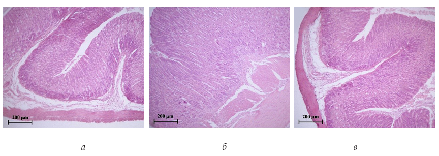
Рис. 1. Участок желудка мышей в группах Cont (а), ContW (б) и ExpChA (в).
Окрашивание гематоксилином и эозином. Увеличение × 100
Fig. 1. Stomach section of mice in the Cont (а), ContW (б), and ExpChA (в) groups.
Hematoxylin and eosin staining. Magnification × 100
В группе животных, получавших ChA (ExpChA), патологических изменений в желудке также не обнаружено (рис. 1, в). Слизистая оболочка оставалась интактной, без эрозий или изъязвлений. Подслизистая основа и мышечные слои не имели признаков отека, фиброза или дистрофии. Железистый аппарат сохранял характерные плотность и морфологию. В собственной пластинке слизистой отмечалась умеренная диффузная полиморфноядерная клеточная инфильтрация без формирования очаговых лимфоидных скоплений. Таким образом, гистологическая картина соответствовала сохранной архитектонике желудочной стенки; обусловленные реакцией стресс или раздражающим действием кислоты эрозивно-язвенные или выраженные воспалительные процессы отсутствовали.
Морфофункциональное состояние печени
Морфологическая оценка печени не выявила патологических отклонений ни в одной из исследованных групп. У интактных животных (Cont) и мышей, получавших воду (ContW), орган имел обычные макроскопические характеристики: стандартные размеры, красно-коричневый цвет, гладкую капсулу без видимых очагов некроза или кровоизлияний. При микроскопии после стандартного окрашивания гематоксилином и эозином паренхима печени была представлена классическими печеночными дольками с сохранной гистоархитектоникой (рис. 2, а, б). Гепатоциты имели центрально расположенные ядра без признаков кариопикноза или кариорексиса, их цитоплазма была гомогенной, без вакуолизации (рис. 2, а, б). Соединительнотканные прослойки по ходу портальных трактов оставались тонкими, без морфологических признаков развития токсического фиброза печени (рис. 2, а, б).
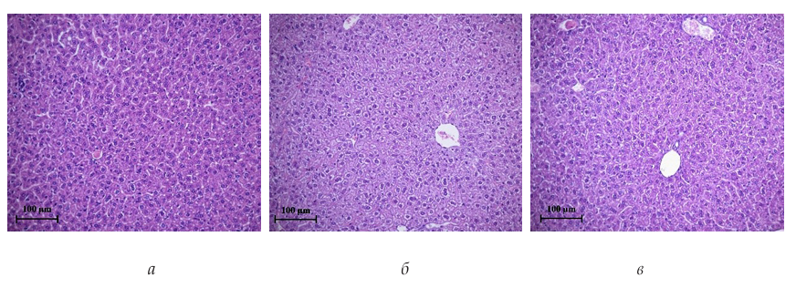
Рис. 2. Участок печени мышей в группах Cont (а), ContW (б) и ExpChA (в).
Окрашивание гематоксилином и эозином. Увеличение × 200
Fig. 2. Liver section of mice in the Cont (а), ContW (б), and ExpChA (в) groups.
Hematoxylin and eosin staining. Magnification × 200
Аналогичная картина наблюдалась в группе, получавшей ChA (ExpChA) (рис. 2, в). Внутрижелудочное введение ChA на протяжении 45 суток не привело к развитию морфологических изменений. В паренхиме печени и в портальных трактах отсутствовали признаки дистрофии, фокального или массивного некроза гепатоцитов, холестаза или воспалительной лейкоцитарной инфильтрации (рис. 2, в). Распределение двуядерных гепатоцитов в дольках было равномерным и соответствовало норме для данного вида животных — менее 30 % [39; 40] — во всех исследуемых группах (табл. 1). Так, в группе интактных животных (Cont) доля двуядерных гепатоцитов составляла 14 (12; 15) % (абсолютное число двуядерных гепатоцитов — 414 (355; 444)), в контроле процедуры введения (ContW) — 13 (10; 15) % (389 (299; 448)), а у мышей, получавших ChA (ExpChA), — 13 (11; 17) % (420 (355; 549)) (табл. 1). При множественном сравнении (Van der Waerden test, p = 0,68, табл. 1) статистически значимых различий между группами не обнаружено.
Таблица 1
Результаты морфометрического анализа цифровых изображений
гистологических срезов печени в исследуемых группах
Table 1
Results of morphometric analysis of digital images of histological sections
of the liver in the study groups
|
Группа |
N |
Доля двуядерных гепатоцитов, % от общего числа гепатоцитов Me (Q1;Q3) |
Множественное сравнение |
Анализ post-hoc |
|
Cont |
10 |
14 (12; 15) |
p = 0,68 |
— |
|
ContW |
10 |
13 (10; 15) |
||
|
ExpChA |
10 |
13 (11; 17) |
Примечание: N — объем выборки (число животных в группе). У каждого животного для подсчетов использовали 10 уникальных полей зрения на увеличении ×200.
Note: N — sample size (number of animals in the group). For each animal, 10 unique fields of view were used for calculations at a magnification of ×200.
Таким образом, 45-дневное пероральное назначение ChA в дозе 10 мг/кг не влияет на регенераторную активность паренхимы печени, оцениваемую по уровню бинуклеации, что позволяет сделать вывод об отсутствии токсического действия ChA на ткань печени в условиях проведенного эксперимента.
Морфофункциональное состояние почек
Проведенное гистологическое исследование почек показало, что структура органа во всех экспериментальных группах соответствовала норме (рис. 3).
У интактных животных (Cont) и мышей контрольной группы, получавших воду (ContW), наблюдалась типичная архитектоника с четкой дифференциацией на корковое и мозговое вещество (рис. 3, а, б, в, г). Почечные тельца были сохранны, клубочки имели нормальную клеточность и форму, капсула Шумлянского — Боумена не была утолщена или деформирована (рис. 3, б, г). Канальцевый аппарат, включая проксимальные и дистальные извитые канальцы, не имел признаков дистрофии, некроза или атрофии. Эпителий канальцев сохранял целостность, а просветы не содержали цилиндров (рис. 3, а, б, в, г). Строма органа была без признаков отека, очаговой лимфоидной инфильтрации или фиброза.
Длительное (45-дневное) внутрижелудочное введение ChA (группа ExpChA) не вызвало визуальных и микроскопических патологических изменений в почечной ткани. Гистологическая картина была полностью сопоставима с таковой в обеих контрольных группах (рис. 3). Как и в контрольных группах, отсутствовали вакуолизация или гиалиново-капельная дистрофия эпителия канальцев, склероз или гиалиноз клубочков, расширение (дилатация) канальцев или интерстициальный отек, воспалительная инфильтрация (рис. 3, д, е). Отсутствие вышеуказанных признаков свидетельствует о том, что ChA не обладает раздражающим или иммунопатологическим эффектом на почечную паренхиму при выбранном режиме введения.
Данные гистологического анализа получили подтверждение в результатах морфометрического исследования. Относительная площадь коркового вещества почки у животных всех групп соответствовала норме для данного вида — 60—80 % [41; 42]. У интактных животных (Cont) она составила 65 (61; 70)%, в контрольной группе (ContW) — 67 (60; 71)%, у мышей, получавших ChA (ExpChA), — 66 (62; 71)%; статистически значимых различий между группами не выявлено (Van der Waerden test, p = 0,72, табл. 2). Удельное количество почечных телец также не имело отличий в сравниваемых группах: 9 (7; 10) шт/ мм2 в группе Cont, 8 (6; 9) шт/мм2 в ContW и 8 (6; 10) шт/мм2 в ExpChA (Van der Waerden test, p = 0,90, табл. 2). Средняя площадь одного почечного тельца составила 1855,85 ± 245,63 мкм2 у интактных животных, 1920,41 ± 198,45 мкм2 в контроле процедуры введения и 2053,20 ± 266,77 мкм2 в опытной группе. При множественном сравнении статистически значимых различий также не обнаружено (Welch’s ANOVA, p = 0,28, табл. 2). Исследуемые характеристики почечных телец также соотносились с нормой [43; 44].
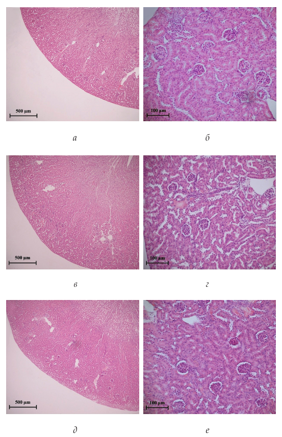
Рис. 3. Участок почек мышей в группах Cont (а, б), ContW (в, г) и ExpChA (д, е).
Окрашивание гематоксилином и эозином. Увеличение × 50 (а, в, д) и ×200 (б, г, е)
Fig. 3. Kidney section of mice in the Cont (а, б), ContW (в, г), and ExpChA (д, е) groups.
Hematoxylin and eosin staining. Magnification × 50 (а, в, д) and ×200 (б, г, е)
Таблица 2
Результаты морфометрического анализа цифровых изображений
гистологических срезов почек в исследуемых группах
Table 2
Results of morphometric analysis of digital images of histological sections
of kidneys in the study groups
|
Группа |
N |
Площадь коркового вещества, % от общей площади почки Me (Q1;Q3) |
Удельное количество почечных телец, шт/мм2 Me (Q1;Q3) |
Средняя площадь одного почечного тельца, мкм2 X ± SD |
|
Cont |
10 |
65 (61; 70) |
9 (7; 10) |
1855,85 ± 245,63 |
|
ContW |
10 |
67 (60; 71) |
8 (6; 9) |
1920,41 ± 198,45 |
|
ExpChA |
10 |
66 (62; 71) |
8 (6; 10) |
2053,20 ± 266,77 |
|
Множественное сравнение |
p = 0,72 |
p = 0,90 |
p = 0,28 |
|
|
Анализ post-hoc |
— |
— |
— |
|
Примечание: N — объем выборки (число животных в группе). Множественное сравнение и анализ post-hoc для признаков «Площадь коркового вещества» и «Удельное количество почечных телец» выполнены с помощью Van der Waerden test и Steel — Dwass test соответственно; для признака «Средняя площадь одного почечного тельца» — с помощью Welch’s ANOVA и Games — Howell test.
Note: N — sample size (number of animals per group). Multiple comparisons and post-hoc analysis for the Cortex area and Specific renal corpuscle count traits were performed using the Van der Waerden test and Steel — Dwass test, respectively; for the Average renal corpuscle area trait — Welch’s ANOVA and Games — Howell test.
Таким образом, 45-суточное внутрижелудочное введение ChA в дозе 10 мг/ кг не влияет на морфофункциональное состояние почек, включая соотношение коркового и мозгового вещества, количество и размеры почечных телец, что свидетельствует об отсутствии у соединения нефротоксического действия в условиях проведенного эксперимента.
Морфофункциональное состояние селезенки
Гистологическое исследование селезенки не выявило наличие патологических изменений, а также качественных и количественных различий в структуре органа ни в одной из исследуемых групп (рис. 4).
У животных контрольных групп (Cont, ContW) наблюдалась типичная гистоархитектоника с четким разделением на белую и красную пульпу (рис. 4, а, г). Лимфоидные фолликулы белой пульпы были хорошо различимы, с заметными герминативными центрами (рис. 4, б, в, д), свидетельствующими об активном иммунном статусе. Кроме того, визуализировались периартериолярные лимфоидные муфты (рис. 4, б, в, е), окружающие ветви центральной артерии. Маргинальная зона не имела признаков расширения или истощения (рис. 4, б, в, д, е). Красная пульпа была представлена типичными синусоидными структурами с умеренным физиологическим кровенаполнением, без признаков застойных явлений, экстрамедуллярного кроветворения или скопления сидерофагов (рис. 4, а, б, в, г, д, е).
У мышей, получавших ChA (группа ExpChA), картина была полностью сопоставима с контролем (рис. 4). Не отмечено изменений в соотношении белой и красной пульпы, а также в структуре лимфоидных образований (рис. 4, ж, з, и). Лимфоидные фолликулы сохраняли высокую клеточность (рис. 4, з, и). Признаков атрофии лимфоидной ткани, гиперплазии или воспалительной инфильтрации в строме органа обнаружено не было (рис. 4, ж). Красная пульпа также не имела отличий от контроля: синусы были умеренно заполнены форменными элементами крови, трабекулы не были утолщены (рис. 4, ж, з, и).
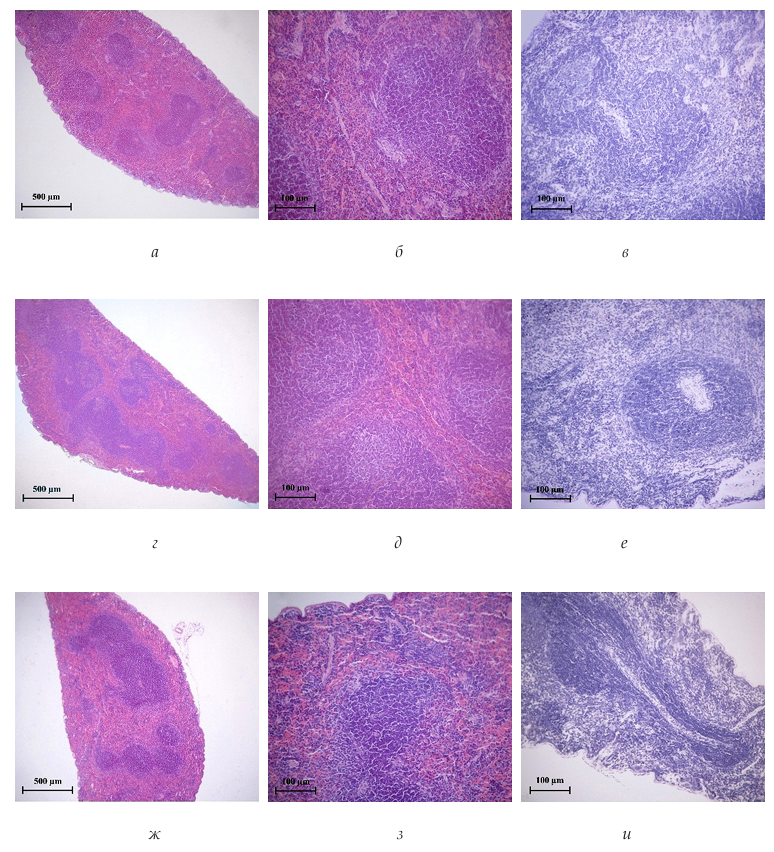
Рис. 4. Участок селезенки мышей в группах Cont (а, б, в), ContW (г, д, е)
и ExpChA (ж, з, и). Окрашивание гематоксилином и эозином (а, б, г, д, ж, з),
и по методу Эйнарсона (в, е, и). Увеличение × 50 (а, г, ж) и × 200 (б, в, д, е, з, и).
Fig. 4. Spleen section of mice in the Cont (а, б, в), ContW (г, д, е)
and ExpChA (ж, з, и) groups. Staining with hematoxylin and eosin (а, б, г, д, ж, з)
and by the Einarson method (в, е, и). Magnification × 50 (а, г, ж) and × 200 (б, в, д, е, з, и).
Результаты морфометрического анализа подтверждают описательные данные. Площадь белой пульпы у интактных животных (Cont) составила 22 (17; 23)%, в контрольной группе (ContW) — 20 (18; 25)%, у мышей, получавших ChA (ExpChA), — 23 (20; 25)%; статистически значимых различий между группами не выявлено (Van der Waerden test, p = 0,85, табл. 3). Соотношение площадей белой и красной пульпы также не имело существенных отличий: 3,55 (3,35; 4,88) в группе Cont, 4,00 (3,00; 4,56) в ContW и 3,35 (3,00; 4,00) в ExpChA (Van der Waerden test, p = 0,67, табл. 3). При этом нормальная относительная площадь красной пульпы и ее отношение к белой для мышей составляют 70—90 % и 4/1 — 2,5/1 соответственно [45—47]. Число лимфоидных фолликулов на срез оставалось сопоставимым во всех трех группах: для интактной группы (Cont) — 10 (9; 13), для животных, получавших воду (ContW), — 11 (10; 13), а ChA (ExpChA) — 10 (8; 11) (Van der Waerden test, p = 0,41, табл. 3).
Таблица 3
Результаты морфометрического анализа цифровых изображений гистологических срезов селезенки в исследуемых группах
Table 3
Results of morphometric analysis of digital images of histological sections
of the spleen in the study groups
|
Группа |
N |
Площадь белой пульпы, % от общей площади среза Me (Q1; Q3) |
Отношение площадей белой и красной пульпы Me (Q1; Q3) |
Число фолликулов на весь срез* Me (Q1; Q3) |
|
Cont |
10 |
22 (17; 23) |
3,55 (3,35; 4,88) |
10 (9; 13) |
|
ContW |
10 |
20 (18; 25) |
4,00 (3,00; 4,56) |
11 (10; 13) |
|
ExpChA |
10 |
23 (20; 25) |
3,35 (3,00; 4,00) |
10 (8; 11) |
|
Множественное сравнение Van der Waerden test |
p = 0,85 |
p = 0,67 |
p = 0,41 |
|
|
Анализ post-hoc Steel — Dwass test |
— |
— |
— |
|
Примечание: N — объем выборки (число животных в группе); * — площади срезов не отличались в исследуемых группах и составляли около 20 мм2.
Note: N — sample size (number of animals in a group). (*) — Section areas did not differ between the study groups and were approximately 20 mm2.
Таким образом, 45-суточное внутрижелудочное введение ChA в дозе 10 мг/кг не влияет на морфофункциональное состояние селезенки, что свидетельствует об отсутствии у тестируемого вещества повреждающего влияния на иммунокомпетентный орган в условиях проведенного эксперимента.
Обсуждение и выводы
ChA представляет значительный интерес как ММ с широким спектром фармакологической активности [10], включающей противовоспалительное [12; 16], противоаллергическое [13; 14], нейропротекторное [17; 18] и регенеративное [20—23] действие. Однако, несмотря на растущее количество данных о ее эффективности, систематические исследования безопасности, особенно при длительном пероральном применении, остаются фрагментарными. В частности, практически отсутствуют сведения о влиянии хронического введения ChA на морфофункциональное состояние органов пищеварения и выделения, что создает пробел в оценке ее общего терапевтического потенциала.
Результаты настоящей работы впервые демонстрируют, что ежедневное внутрижелудочное введение ChA в концентрации 10 мг/кг в течение 45 суток не вызывает патологических структурных изменений в ключевых органах-мишенях (желудок, печень, почки) и не нарушает гистоархитектонику селезенки у мышей линии Balb/c.
Пероральный путь введения рассматривается как наиболее предпочтительный для терапевтического применения ММ, что обусловлено простотой применения, хорошей биодоступностью и предсказуемой фармакокинетикой [4; 5]. Вместе с тем желудочно-кишечный тракт (ЖКТ), будучи первым барьером на пути ксенобиотика и обеспечивая не только транспорт, но и расщепление, переваривание и всасывание нутриентов, электролитов и воды, особенно уязвим к повреждающему действию лекарственных средств [48]. Данная проблема наиболее актуальна для соединений с противовоспалительной и анальгетической активностью, поскольку многие нестероидные противовоспалительные препараты ассоциированы с развитием гастропатий, эрозий и язв слизистой оболочки желудка [49].
ChA, согласно литературным данным, обладает фармакологическим профилем, характерным для анальгетиков и противовоспалительных средств [11; 16]. Также следует принимать во внимание раздражающее действие органической кислоты. Это делает оценку ее потенциального влияния на состояние желудка достаточно важной для определения безопасности перорального применения.
Существующие сведения о переносимости ChA со стороны ЖКТ ограничены и носят фрагментарный характер. В частности, имеющиеся данные о безопасности внутрижелудочного введения вещества получены в краткосрочных экспериментах (до 75 мин) и носят скорее описательный характер в рамках исследований, сфокусированных на синтезе и первичной фармакологической оценке производных 4(1H)-пиридинона, к которым относится ChA [16].
Результаты проведенного гистологического анализа свидетельствуют о том, что длительное введение ChA не вызывало раздражения или повреждения слизистой оболочки желудка, а также не приводило к развитию воспалительных процессов в слизистом, подслизистом или мышечном слоях (см. рис. 1, в). Общая морфологическая картина соответствовала норме, что согласуется с имеющимися данными об отсутствии острых неблагоприятных реакций со стороны желудка после введения ChA [16]. Более того, полученные данные коррелируют с результатами исследований, демонстрирующих защитную роль ChA в модели язвенного колита, индуцированного декстрансульфатом натрия [11]. Можно предположить, что собственные противовоспалительные свойства ChA могут компенсировать потенциально раздражающее воздействие при ее хроническом контакте со слизистой оболочкой ЖКТ.
Печень играет центральную роль в метаболизме ксенобиотиков, выступая в качестве основного органа детоксикации благодаря наличию специализированных ферментативных систем [50]. Эти системы включают комплекс ксенобиотик-биотрансформирующих ферментов I фазы (окисление, восстановление, гидролиз) и II фазы (конъюгация), а также транспортные белки III фазы, ответственные за выведение метаболитов [51; 52]. Благодаря такой многоуровневой организации печень способна как к деактивации потенциально опасных соединений [50; 53], так и, в некоторых случаях, к их метаболической активации в более реакционноспособные формы [53; 54].
Именно поэтому оценка состояния печени является критически важным элементом скрининга любых новых биологически активных веществ, поскольку этот орган из-за своей функциональной нагрузки часто становится основной мишенью для гепатотоксических эффектов [55]. Существующие данные о безопасности ChA для печеночной паренхимы носят ограниченный, но обнадеживающий характер. Публикации свидетельствуют об отсутствии признаков острой токсичности даже при введении высоких доз ChA (до 2000 мг/кг) в течение 14 дней, а также о хорошей переносимости при ее длительном применении в более низких дозах (до 40 мг/кг) на крысиных моделях [56].
Результаты настоящего исследования, в котором проводилась гистологическая оценка состояния печени после 45-дневного ежедневного внутрижелудочного введения ChA в дозе 10 мг/кг, полностью согласуются с этими данными. Проведенный анализ не выявил каких-либо патологических отклонений от нормы (см. рис. 2, табл. 1). Отсутствие морфологических изменений в печени в нашем эксперименте, дополняя ранее полученные сведения о переносимости, укрепляет вывод о благоприятном профиле безопасности ChA и отсутствии у нее выраженного гепатотоксического потенциала в изученном режиме дозирования.
В метаболизме и элиминации ксенобиотиков наряду с печенью активное участие принимают почки [57]. Этот орган обладает собственным спектром специфических ферментов, способных к биотрансформации лекарственных веществ, а также играет ключевую роль в их окончательном выведении из организма [58; 59]. Высокая уязвимость почек к лекарственной токсичности обусловлена рядом физиологических особенностей: интенсивным кровотоком, значительным потреблением кислорода, концентрирующей функцией, активными процессами биотрансформации и выраженной активностью мембранных транспортных систем [60; 61]. Хотя для выявления повреждения почек традиционно используются биохимические маркеры в моче [62—64], гистологическое исследование предоставляет наиболее полную информацию, позволяя непосредственно оценить структурную целостность и морфологию почечной ткани [62; 65]. Учитывая определенную, хотя и ограниченную водорастворимость ChA, можно говорить о том, что ее выведение предположительно осуществляется почечным путем, что делает оценку состояния почек критически важной.
Проведенные в настоящей работе гистологические и морфометрические исследования показали, что 45-суточное введение ChA в дозе 10 мг/кг не приводит к развитию нефропатии, воспалительной инфильтрации или дистрофических изменений в почечной ткани (см. рис. 3). Количественный морфометрический анализ также не выявил статистически значимых отличий от контрольных групп по таким параметрам, как относительная площадь коркового и мозгового вещества, количество, а также средняя площадь почечных телец (см. табл. 2). Эти данные согласуются с результатами предыдущих исследований, в которых не было обнаружено признаков острой и субхронической токсичности ChA даже при использовании значительно более высоких доз (до 2000 мг/кг) [56]. Косвенным свидетельством вовлеченности почек в метаболизм ChA являются наши данные о возможном взаимодействии этого соединения с транспортером URAT1 (human urate transporter 1) [66], преимущественно экспрессирующимся в почках [67] и участвующим в реабсорбции мочевой кислоты [68].
Наличие такого потенциального механизма транспорта поднимает вопрос о возможном влиянии ChA на почечную функцию, которое теоретически могло быть как позитивным (например, за счет конкурентного ингибирования URAT1), так и негативным (риск нефропатии или изменения экскреторных процессов). Хотя настоящее исследование не ставило целью прямое подтверждение участия URAT1 в транспорте ChA, полученные морфологические данные об отсутствии повреждений свидетельствуют в пользу благоприятного профиля. Более того, имеющиеся литературные данные о способности ChA снижать нефротоксичность, индуцированную цисплатином, за счет модуляции воспалительных цитокинов [69] позволяют предположить, что ее влияние на почки при длительном введении может носить нейтральный или потенциально протекторный характер.
Хотя селезенка не участвует напрямую в метаболизме ксенобиотиков, наличие у ChA выраженных иммуномодулирующих свойств, описанных в литературе, побудило нас включить оценку ее гистоархитектоники в данную работу. Проведенное исследование показало, что курсовое (45 суток) внутрижелудочное введение ChA в дозе 10 мг/кг не привело к каким-либо структурным изменениям в селезенке. Не было отмечено статистически значимых отличий в соотношении белой и красной пульпы (см. табл. 3), а также в структурной организации лимфоидных образований (см. рис. 4). Лимфоидные фолликулы сохраняли клеточную плотность и архитектонику (см. рис. 4). В строме органа отсутствовали признаки атрофии лимфоидной ткани, гиперплазии или очаговой воспалительной инфильтрации, что свидетельствует об отсутствии морфологически выраженного воздействия на иммунный орган при систематическом введении соединения.
Интересно, что отсутствие структурных изменений на тканевом уровне контрастирует с данными, демонстрирующими четкое функциональное иммуномодулирующее действие ChA в других экспериментальных моделях. Так, в исследованиях на мышах с аллергическим ринитом применение ChA приводило к значимому снижению патологической реакции на аллергены, что коррелировало с уменьшением количества эозинофилов и тучных клеток в слизистых оболочках [13]. В другой работе на крысах внутрижелудочное введение ChA вызывало снижение тканевой эозинофильной инфильтрации, уменьшение количества циркулирующих эозинофилов и уровня сывороточного IgE, что в конечном итоге снижало смертность после иммунизации [14]. Таким образом, полученные нами данные позволяют предположить, что иммуномодулирующий эффект ChA, по-видимому, реализуется на уровне регуляции конкретных клеточных популяций и гуморальных факторов без явных морфологических изменений, что может рассматриваться как благоприятный признак селективности ее действия.
Важно отметить, что данное исследование имеет определенные ограничения. В частности, работа проводилась с использованием только одной, достаточно низкой, но активной дозировки (10 мг/кг) и на одной модели животных — самцах мышей линии Balb/c. Это не позволяет судить о возможных дозозависимых эффектах ChA и экстраполировать полученные данные на другие виды млекопитающих. Тем не менее гистологические методы проведенного анализа позволили получить объективные и репрезентативные доклинические результаты для данной экспериментальной модели.
Заключение
В данном исследовании впервые проведена комплексная оценка влияния длительного (45 суток) внутрижелудочного введения ChA на морфофункциональное состояние ключевых органов, участвующих в метаболизме и экскреции ксенобиотиков (желудок, печень, почки), а также иммунокомпетентного органа (селезенка). Полученные результаты демонстрируют отсутствие патологических структурных изменений во всех исследуемых органах при использовании вещества в концентрации 10 мг/кг. При этом ChA и ее кальциевая соль в указанной дозе обладают специфической (остеогенной) активностью в отношении мезенхимных стромальных / стволовых клеток как in vitro, так и in vivo [20; 21].
Полученные данные свидетельствуют об отсутствии общетоксического действия низкой концентрации вещества на линейных мышах, что открывает хорошие перспективы для дальнейших испытаний для регистрации активной фармацевтической субстанции полностью отечественного происхождения, имеющей существенное значение для развития арсенала регенеративной биомедицины, в первую очередь для биоинженерии костной ткани.
Reference
- Mitchell J., Lo K. W. H. Small molecule-mediated regenerative engineering for craniofacial and dentoalveolar bone. Front. Bioeng. Biotechnol. 2022, 10, 1003936, DOI: 10.3389/fbioe.2022.1003936.
- Lipinski C. A., Lombardo F., Dominy B. W., Feeney P. J. Experimental and computational approaches to estimate solubility and permeability in drug discovery and development settings. Adv. Drug. Deliv. Rev. 2001, 46(1—3), 3—26, DOI: 10.1016/s0169-409x(00)00129-0.
- Scott D. E., Bayly A. R., Abell C., Skidmore J. Small molecules, big targets: drug discovery faces the protein-protein interaction challenge. Nat. Rev. Drug. Discov. 2016, 15(8), 533—550, DOI: 10.1038/nrd.2016.29.
- Chen S., Song Z., Zhang A. Small-Molecule Immuno-Oncology Therapy: Advances, Challenges and New Directions. Curr. Top. Med. Chem. 2019, 19(3), 180—185, DOI: 10.2174/1568026619666190308131805.
- Toogood P. L. Small molecule immuno-oncology therapeutic agents. Bioorg. Med. Chem. Lett. 2018, 28(3), 319—329, DOI: 10.1016/j.bmcl.2017.12.044.
- Morgan S., Grootendorst P., Lexchin J., Cunningham C., Greyson D. The cost of drug development: a systematic review. Health Policy. 2011, 100(1), 4—17, DOI: 10.1016/j.healthpol.2010.12.002.
- Newman D. J., Cragg G. M. Natural Products as Sources of New Drugs over the Nearly Four Decades from 01/1981 to 09/2019. J. Nat. Prod. 2020, 83(3), 770—803, DOI: 10.1021/acs.jnatprod.9b01285.
- Nielsen H. M., Jorgensen L. Challenges in Delivery of Biopharmaceuticals; the Need for Advanced Delivery Systems. Delivery Technologies for Biopharmaceuticals, John Wiley & Sons, Ltd; 2009. 1—8, DOI: 10.1002/9780470688397.ch1.
- Makurvet F. D. Biologics vs. small molecules: Drug costs and patient access. Medicine in Drug Discovery. 2021, 9, 100075, DOI: 10.1016/j.medidd.2020.100075.
- Miroshnichenko L. A., Polyakova T. Y., Avdeeva E. Y., Krivoshchekov S. V., Khlusov I. A., Belousov M. V. Chelidonic Acid and its Derivates: General Spectrum of Biological Activity and Osteogenic Properties (Review). Drug development & registration. 2022, 11(4), 60—71, DOI: 10.33380/2305-2066-2022-11-4-60-71.
- Kim D.-S., Kim S.-J., Kim M.-C., Jeon Y.-D., Um J., Hong S.-H. The therapeutic effect of chelidonic acid on ulcerative colitis. Biol. Pharm. Bull. 2012, 35(5), 666—671, DOI: 10.1248/bpb.35.666.
- Liu H., Zheng Y., Li C., Zheng Y., Wang D., Wu Z., et al. Discovery of Anti-inflammatory Ingredients in Chinese Herbal Formula Kouyanqing Granule based on Relevance Analysis between Chemical Characters and Biological Effects. Sci. Rep. 2015, 5, 18080, DOI: 10.1038/srep18080.
- Oh H.-A., Kim H.-M., Jeong H.-J. Beneficial effects of chelidonic acid on a model of allergic rhinitis. Int. Immunopharmacol. 2011, 11(1), 39—45, DOI: 10.1016/j.intimp.2010.10.002.
- Singh D. K., Gulati K., Ray A. Effects of chelidonic acid, a secondary plant metabolite, on mast cell degranulation and adaptive immunity in rats. Int. Immunopharmacol. 2016, 40, 229—234, DOI: 10.1016/j.intimp.2016.08.009.
- Aytemir M. D., Uzbay T., Erol D. D. New 4(1H)-pyridinone derivatives as analgesic agents. Arzneimittelforschung. 1999, 49(3), 250—254, DOI: 10.1055/s-0031-1300409.
- Oztürk G., Erol D. D., Aytemir M. D., Uzbay T. New analgesic and antiinflammatory agents 4(1H)-pyridinone derivatives. Eur. J. Med. Chem. 2002, 37(10), 829—834, DOI: 10.1016/s0223-5234(02)01390-9.
- Jeong H.-J., Yang S.-Y., Kim H.-Y., Kim N.-R., Jang J.-B., Kim H.-M. Chelidonic acid evokes antidepressant-like effect through the up-regulation of BDNF in forced swimming test. Exp. Biol. Med. (Maywood). 2016, 241(14), 1559—1567, DOI: 10.1177/1535370216642044.
- Hamada Y., Suzuki K., Nakanishi T., Sarma D., Ohta H., Yamaguchi R., et al. Structure-activity relationship study of BACE1 inhibitors possessing a chelidonic or 2,6-pyridinedicarboxylic scaffold at the P(2) position. Bioorg. Med. Chem. Lett. 2014, 24(2), 618—623, DOI: 10.1016/j.bmcl.2013.12.007.
- Porter T. G., Martin D. L. Chelidonic acid and other conformationally restricted substrate analogues as inhibitors of rat brain glutamate decarboxylase. Biochem. Pharmacol. 1985, 34(23), 4145—4150, DOI: 10.1016/0006-2952(85)90207-2.
- Avdeeva E., Shults E., Rybalova T., Reshetov Y., Porokhova E., Sukhodolo I., et al. Chelidonic Acid and Its Derivatives from Saussurea Controversa: Isolation, Structural Elucidation and Influence on the Osteogenic Differentiation of Multipotent Mesenchymal Stromal Cells In vitro. Biomolecules. 2019, 9(5), 189, DOI: 10.3390/biom9050189.
- Avdeeva E., Porokhova E., Khlusov I., Rybalova T., Shults E., Litvinova L., et al. Calcium Chelidonate: Semi-Synthesis, Crystallography, and Osteoinductive Activity In vitro and In vivo. Pharmaceuticals (Basel). 2021, 14(6), 579, DOI: 10.3390/ph14060579.
- Nasibov T., Gorokhova A., Starosvetskaya A., Bariev U., Nosov V., Litvinova L., et al. In situ and in silico modeling of the hematopoiesis-inducing effect of chelidonic acid. Russ. J. Immunol. 2024, 27(2), 187—196, DOI: 10.46235/1028-7221-16768-ISA.
- Nasibov T., Gorokhova A., Porokhova E., Shupletsova V., Yurova K., Avdeeva E., et al. A hypothesis of mesenchymal stem cell osteogenic differentiation mediated by chelidonic acid through the calcium import: original research and computer simulation. Histochem. Cell Biol. 2024, 163(1), 14, DOI: 10.1007/s00418-024-02342-5.
- Nasibov T. F., Gorokhova A. V., Brazovsky K. S., Porokhova E. D., Avdeeva E. Y., Belousov M. V., et al. The pipeline for in silico prediction of target genes and target metabolic pathways for small molecules, using chelidonic acid as an example. Adv. Target Biomed. 2025, 1(1), 6—19, DOI: 10.5922/ATB-2025-1-1-1.
- Pearse A. G. E. Histochemistry, Theoretical and Applied. London: Churchill Livingstone, 1968. 759.
- Shakhov V. P., Khlusov I. A., Dambaev K. V., Zaitsev K. V., Salmina A. B., Shakhova S. S. et al. Introduction to methods of cell culture, bio-engineering of organs and tissues. Tomsk: STT, 2004. 385.
- Royston P. Remark AS R94: a remark on algorithm AS 181: the W-test for normality. J. R. Stat. Soc. Ser. C Appl. Stat. 1995, 44(4), 547–551, DOI: 10.2307/2986146.
- Celik N. Welch’s ANOVA: Heteroskedastic skew-t error terms: Communications in Statistics. Theory and Methods. 2022, 51(9), 3065—3076, DOI: 10.1080/03610926.2020.1788084.
- Delacre M., Lakens D., Leys C. Why Psychologists Should by Default Use Welch’s t-test Instead of Student’s t-test. International Review of Social Psychology. 2017, 30(1), 92, DOI: 10.5334/irsp.82.
- Tomarken A. J., Serlin R. C. Comparison of ANOVA alternatives under variance heterogeneity and specific noncentrality structures. Psychol. Bull. 1986, 99(1), 90—99, DOI: 10.1037/0033-2909.99.1.90.
- Levy K. J. An empirical comparison of the ANOVA F-test with alternatives which are more robust against heterogeneity of variance. J. Stat. Comput. Sim. 1978, 8(1), 49—57, DOI: 10.1080/00949657808810247.
- Lix L. M., Keselman J. C., Keselman H. J. Consequences of Assumption Violations Revisited: A Quantitative Review of Alternatives to the One-Way Analysis of Variance F Test. Review of Educational Research. 1996, 66(4), 579—619, DOI: 10.3102/00346543066004579.
- Tamhane A. C. A Comparison of Procedures for Multiple Comparisons of Means with Unequal Variances. J. Am. Stat. Assoc. 1979, 74(367), 471—480, DOI: 10.1080/01621459.1979.10482541.
- Elamir E. On uses of van der Waerden test: a graphical approach. 2022. Available from: arXiv: 2203.02148, DOI: 10.48550/arXiv.2203.02148.
- Macunluoglu A. C., Ocakoğlu G. Comparison of the performances of non-parametric k-sample test procedures as an alternative to one-way analysis of variance. Eur. Res. J. 2023, 9, 687—696, DOI: 10.18621/eurj.1037546.
- Luepsen H. Comparison of nonparametric analysis of variance methods: a Monte Carlo study — a vote for van der Waerden. Commun. Stat. Simul. Comput. 2018, 47(9), 2547—2576, DOI: 10.1080/03610918.2017.1353613.
- Douglas C. E., Michael F. A. On distribution-free multiple comparisons in the one-way analysis of variance. Communications in Statistics - Theory and Methods. 1991, 20(1), 127—139, DOI: 10.1080/03610929108830487.
- Miller R. G. Nonparametric Techniques. In: Miller RG, editor. Simultaneous Statistical Inference, N Y: Springer, 1981. 129—188, DOI: 10.1007/978-1-4613-8122-8_4.
- Madra S., Styles J., Smith A. G. Perturbation of hepatocyte nuclear populations induced by iron and polychlorinated biphenyls in C57BL/10ScSn mice during carcinogenesis. Carcinogenesis. 1995, 16(4), 719—727, DOI: 10.1093/carcin/16.4.719.
- Jácome F. C., Caldas G. C., Rasinhas A. C., de Almeida A. L. T., de Souza D. D. C., Paulino A. C., et al. Comparative analysis of liver involvement caused by two DENV-2 lineages using an immunocompetent murine model. Sci. Rep. 2021, 11(1), 9723, DOI: 10.1038/s41598-021-88502-2.
- Missbach-Guentner J., Pinkert-Leetsch D., Dullin C., Ufartes R., Hornung D., Tampe B., et al. 3D virtual histology of murine kidneys -high resolution visualization of pathological alterations by micro computed tomography. Sci. Rep. 2018, 8, 1407, DOI: 10.1038/s41598-018-19773-5.
- Sadeghinezhad J., Nyengaard J. R. Oxaliplatin Treatment for 3-Weeks Does not Affect Mice Kidney Structure: A Stereological Study. Iranian Journal of Veterinary Medicine. 2022, 16(2), 101—119, DOI: 10.22059/IJVM.2021.311503.1005132.
- Baldelomar E. J., Charlton J. R., Beeman S. C., Hann B. D., Cullen-McEwen L., Pearl V. M., et al. Phenotyping by magnetic resonance imaging nondestructively measures glomerular number and volume distribution in mice with and without nephron reduction. Kidney Int. 2016, 89(2), 498—505, DOI: 10.1038/ki.2015.316.
- Sato F., Tsuchihashi S., Kawashima N. Age changes in number and size of the murine renal glomeruli. Exp. Gerontol. 1975, 10, 325–331, DOI: 10.1016/0531-5565(75)90022-4.
- Pislyagin E. A., Manzhulo I. V., Dmitrenok P. S., Aminin D. L. Cucumarioside A2-2 causes changes in the morphology and proliferative activity in mouse spleen. Acta Histochem. 2016, 118(4), 387—392, DOI: 10.1016/j.acthis.2016.03.009.
- Meyer E. M., Schlake W., Grundmann E. Comparative histologic, histochemical, and histomorphometric studies of T and B cell areas in peripheral lymphoid organs of normal young adult BALB/C mice. Pathol. Res. Pract. 1979, 164(2), 141—156, DOI: 10.1016/s0344-0338(79)80019-9.
- Cherkasova M. N., Zhukhovitskii V. G., Borovaya T. G. Comparative Characteristics of the Spleen White Pulp in Experimental Models of Sepsis Caused by Different Strains of Pseudomonas aeruginosa. Bull. Exp. Biol. Med. 2020, 170(1), 46—48, DOI: 10.1007/s10517-020-05001-6.
- Harrison A. P., Erlwanger K. H., Elbrønd V. S., Andersen N. K., Unmack M. A. Gastrointestinal-tract models and techniques for use in safety pharmacology. J. Pharmacol. Toxicol. Methods. 2004, 49(3), 187—199, DOI: 10.1016/j.vascn.2004.02.008.
- Pusztaszeri M. P., Genta R. M., Cryer B. L. Drug-induced injury in the gastrointestinal tract: clinical and pathologic considerations. Nat. Clin. Pract Gastroenterol. Hepatol. 2007, 4, 442—453, DOI: 10.1038/ncpgasthep0896.
- Remmer H. The role of theliver in drug metabolism. Am. J. Med. 1970, 49(5), 617—629, DOI: 10.1016/s0002-9343(70)80129-2.
- Xu C., Li C. Y.-T., Kong A.-N. T. Induction of phase I, II and III drug metabolism/transport by xenobiotics. Arch. Pharm. Res. 2005, 28(3), 249—268, DOI: 10.1007/BF02977789.
- Grant D. M. Detoxification pathways in the liver. J. Inherit. Metab. Dis. 1991, 14(4), 421—430, DOI: 10.1007/BF01797915.
- Apte U., Krishnamurthy P. Detoxification functions of the liver. In: Monga SPS, editor. Molecular pathology of liver diseases. Berlin: Springer; 2011. 147—463, DOI: 10.1007/978-1-4419-7107-4_11.
- You L. Steroid hormone biotransformation and xenobiotic induction of hepatic steroid metabolizing enzymes. Chem. Biol. Interact. 2004, 147(3), 233—246, DOI: 10.1016/j.cbi.2004.01.006.
- Kleiner D. E. The histopathological evaluation of drug-induced liver injury. Histopathology. 2017, 70(1), 81—93, DOI: 10.1111/his.13082.
- Khairnar S. I., Kulkarni Y. A., Murugesan S., Singh K. Effects of Acute and Repeated Dose Toxicity Profiling of Chelidonic Acid in Rats: in Silico and in vivo Evidence. Chem. Biodivers. 2023, 20(12), e202301241, DOI: 10.1002/cbdv.202301241.
- Lohr J. W., Willsky G. R., Acara M. A. Renal drug metabolism. Pharmacol. Rev. 1998, 50(1), 107—141.
- Miners J. O., Yang X., Knights K. M., Zhang L. The Role of the Kidney in Drug Elimination: Transport, Metabolism, and the Impact of Kidney Disease on Drug Clearance. Clin. Pharmacol. Ther. 2017, 102(3), 436—449, DOI: 10.1002/cpt.757.
- Knights K. M., Rowland A., Miners J. O. Renal drug metabolism in humans: the potential for drug-endobiotic interactions involving cytochrome P450 (CYP) and UDP-glucuronosyltransferase (UGT). Br. J. Clin. Pharmacol. 2013, 76(4), 587—602, DOI: 10.1111/bcp.12086.
- Radi Z. A. Kidney Pathophysiology, Toxicology, and Drug-Induced Injury in Drug Development. Int. J. Toxicol. 2019, 38(3), 215—227, DOI: 10.1177/1091581819831701.
- Kim S. Y., Moon A. Drug-induced nephrotoxicity and its biomarkers. Biomol. Ther. (Seoul). 2012, 20(3), 268—272, DOI: 10.4062/biomolther.2012.20.3.268.
- Fuchs T. C., Hewitt P. Biomarkers for drug-induced renal damage and nephrotoxicity-an overview for applied toxicology. AAPS J. 2011, 13(4), 615—631, DOI: 10.1208/s12248-011-9301-x.
- Troth S. P., Simutis F., Friedman G. S., Todd S., Sistare F. D. Kidney Safety Assessment: Current Practices in Drug Development. Semin. Nephrol. 2019, 39(2), 120—131, DOI: 10.1016/j.semnephrol.2018.12.002.
- Griffin B. R., Faubel S., Edelstein C. L. Biomarkers of Drug-Induced Kidney Toxicity. Therapeutic Drug Monitoring. 2019, 41(2), 213—226, DOI: 10.1097/FTD.0000000000000589.
- Huang J. X., Blaskovich M. A., Cooper M. A. Cell- and biomarker-based assays for predicting nephrotoxicity. Expert Opin. Drug Metab. Toxicol. 2014, 10(12), 1621—1635, DOI: 10.1517/17425255.2014.967681.
- Nasibov T., Gorokhova A., Brazovsky K., Ryzhkova A., Porokhova E., Avdeeva E., et al. Potential Transmembrane Proteins-transporters of Chelidonic Acid for its Intracellular Uptake: In Silico Simulation. Pharm. Res. 2025, 42(10), 1749—1773, DOI: 10.1007/s11095-025-03934-z.
- Szabo L., Morey R., Palpant N. J., Wang P. L., Afari N., Jiang C., et al. Statistically based splicing detection reveals neural enrichment and tissue-specific induction of circular RNA during human fetal development. Genome Biol. 2015, 16(1), 126, DOI: 10.1186/s13059-015-0690-5.
- Dai Y., Lee C.-H. Transport mechanism and structural pharmacology of human urate transporter URAT1. Cell Res. 2024, 34, 776—787, DOI: 10.1038/s41422-024-01023-1.
- Khairnar S. I., Kulkarni Y. A., Singh K. Mitigation of cisplatin-induced nephrotoxicity by chelidonic acid in Wistar rats. J. Trace Elem. Med. Biol. 2024, 81, 127321, DOI: j.jtemb.2023.127321.