
Taxonomic composition of the oral microbiome and its relationship with immune parameters in periodontal inflammatory disease
- DOI
- 10.5922/ATB-2026-2-1-8
- Pages
- 124-136
- To cite
- Gimranova I. A., Akmalova G. M., Gareev E. M., Gritsenko V. A. Taxonomic composition of the oral microbiome and its relationship with immune parameters in periodontal inflammatory disease. Advanced targets in Biomedicine. 2026;2(1):124—136. https://doi.org/10.5922/ATB-2026-2-1-8
Abstract
Elucidating the role of periodontopathogenic microorganisms in inflammatory periodontal diseases and the immune response they trigger in the pathogenesis may open up new opportunities for the diagnosis, prevention, and treatment of this pathology.
The aim is to analyze the relationships between the taxonomic features of the microbiome of the gingival sulcus/periodontal pocket contents and immunological indicators in patients with gingivitis and chronic generalized periodontitis (CGP) of varying severity.
Materials and methods. The 16S rRNA gene was sequenced according to the Illumina protocol to determine the species composition of the microbiome in the gingival sulcus and periodontal pocket of 84 patients. The concentration of pro- and anti-inflammatory cytokines was determined in the selected samples using enzyme-linked immunosorbent assay. The peripheral blood of the patients was evaluated for the content of immunoglobulins A, M, and G and the phagocytic activity of neutrophils. Statistical data processing and correlation analysis were performed using the Statistica 10 software package.
Results and discussion. It was found that patients with inflammatory periodontal diseases had increased levels of IL-2, TNFα, and IL-4 in their oral fluid, as well as sIgA, while there was a statistically significant decrease in the levels of IL-6 and IL-10. In the peripheral blood of patients with CGP, there was a decrease in the phagocytic activity of leukocytes and dysgammaglobulinemia. A significant “positive” correlation was found between the presence of representatives of the V. dispar species in the oral microbiome and increased levels of TNFα, IL-2, IL-4, and sIgA (τ = 0.27...0.36, p < 0.05), as well as a negative correlation between the occurrence of F. nucleatum, T. forsythia, and F. alocis with a number of immunological parameters — TNFα, IL-2, IL-4, sIgA, IgA, IgM, IgG (τ = – 0.57... – 0.86, p < 0.01).
Conclusion. The presence of P. gingivalis, F. nucleatum, F. alocis, and V. dispar in the oral microbiome of patients with chronic generalized periodontitis contributes to the suppression of cellular and humoral immunity and is a potential biomarker of severe and progressive disease, which should be taken into account during comprehensive therapy.
Full text
Introduction
Inflammatory periodontal diseases (IPD) (gingivitis, periodontitis) are among the most prevalent diseases of the maxillofacial region, which, as they progress, lead to increased tooth mobility and tooth loss, thereby adversely affecting patients’ quality of life [1; 2]. The recurrence and generalization of this pathology are driven by a chronic, indolent, and poorly controllable infectious-inflammatory process in periodontal tissues. A key role in the etiology and pathogenesis of these diseases is played by periodontopathogenic microorganisms and their interaction with humoral and cellular factors of the host immune system [3—5].
The human oral microbiota/microbiome comprises more than 700 species of various microorganisms that form a symbiotic biofilm on tooth surfaces [6—8]. Dysbiotic shifts in the oral microbiota, arising from the action of various adverse factors (poor oral hygiene, improper dietary habits, stress exposure, etc.) and accompanied by an increase in the pool of periodontopathogens, contribute to the development and progression of inflammatory periodontal diseases, with gingivitis transitioning into chronic generalized periodontitis [2].
Periodontopathogens include Porphyromonas gingivalis, Aggregatibacter actinomycetemcomitans, Tannerella forsythia, Treponema denticola, Fusobacterium nucleatum, Prevotella intermedia, Streptococcus intermedius, Campylobacter rectus, Peptostreptococcus micros, members of the phylum Spirochaetes, and others. All periodontopathogens are divided into two orders: Order I includes P. gingivalis, A. actinomycetemcomitans, and T. forsythia, which promote rapid disease progression, as their pathogenicity/virulence factors are implicated in tissue destruction and intracellular parasitism by the pathogens [9]; Order II comprises T. denticola, F. nucleatum, P. intermedia, S. intermedius, and others, which play a substantial but less critical role in the development of inflammatory periodontal pathology. However, representatives of Order II can form associations with other periodontopathogens, which may subsequently lead to inflammation. Such associations may involve P. gingivalis and T. forsythia, which can initiate an inflammatory process in the subgingival area, while consortia of P. intermedia with other periodontopathogens are linked to disease progression [10; 11].
Furthermore, in addition to the complex of dysbiotic shifts in the structure of the oral microbiota, pathological changes in the immune status make a substantial contribution to the development of IPD [12—14], correlating with the severity of gingivitis and periodontitis [15]. Immune cells stimulated by subgingival plaque extensively infiltrate the gingival connective tissue and secrete numerous cytokines, which play a key role in the additional recruitment of immunocompetent cells, modulation of osteoclast activity, and control of pathogenic microorganisms [16]. Pro-inflammatory cytokines such as IL-1β and the chemokine CXCL6 exert a pronounced regulatory influence on the immune cell response and the inflammatory process associated with the destruction and resorption of hard periodontal tissues [7; 17]. The involvement of various components of adaptive immunity in the pathogenesis of IPD should be emphasized. Thus, in an experimental model of periodontitis in rats, a decrease in cellular (total T-lymphocytes, T-helper cells) and humoral (B-lymphocytes) immunity parameters was observed [18].
Periodontopathogenic microorganisms cause inflammation and destruction in periodontal tissues either directly, by disintegrating the corresponding histostructures of the affected tooth, or indirectly, when, in response to their aggression, a complex of immunopathological mechanisms is triggered, promoting the destruction of alveolar bone, which is accompanied by changes in the microbiota composition and the growth of periodontopathogens [19]. This creates a self-sustaining vicious cycle that maintains and drives the progression of periodontal disease. The literature contains relatively few data on the correlation between the occurrence of certain microbial species and changes in immune status indicators that reflect the progression of periodontitis [20; 21]. Moreover, the focus of such studies is typically limited to a narrow range of known periodontopathogens, whereas the relationships of other representatives of the oral microbiota with the human immune system in IPD remain poorly understood.
Aim of the study
To analyze the relationships between the taxonomic features of the microbiome of the gingival sulcus / periodontal pocket content and immunological indicators in patients with gingivitis and chronic generalized periodontitis of varying severity.
Materials and methods
The study analyzed the microbiome composition of the gingival sulcus and periodontal pocket content and a range of immunological parameters in 84 patients who sought dental treatment at the Primadent Dental Clinic in Ufa between 2021 and 2023. Patients with clinically confirmed diagnoses of inflammatory periodontal diseases were divided into 4 groups: 15 patients with gingivitis (mean age 31.4 years); 30 patients with mild chronic generalized periodontitis (CGP) (mean age 49.8 years); 39 patients with moderate CGP (mean age 53.4 years); the comparison group (control) consisted of 21 healthy individuals without signs of IPD (29.1 years). Patients were excluded from the study if they had a history of systemic diseases; acute or chronic somatic diseases in the decompensation stage; oncological diseases; HIV infection; hepatitis B and C; antibiotic, hormonal, or cytostatic drug intake within 4 months prior to the study; pregnancy; breastfeeding; or smoking. Informed consent was obtained from each participant, ensuring the voluntary and informed nature of their participation in the study. This approach ensured the representativeness of the sample and minimized the influence of extraneous factors on the study results.
Biomaterial sampling was performed by a periodontist by inserting sterile paper endodontic absorbent points, size № 20 according to ISO (META BIOMED, South Korea), into the depth of the gingival sulcus or periodontal pocket for 15 seconds. The points were then placed into sterile tubes containing transport medium (DNA/RNA Shield, Zymo Research, USA). Subsequently, the obtained material was delivered to the laboratory, where DNA was extracted from samples pre-homogenized in a lysis solution with beads using the sorbent column method (QIAamp DNA Mini Kit, Qiagen, Germantown, MD, USA) in accordance with the manufacturer’s recommendations. Sequencing was performed at the Serbalab genetic laboratory (St. Petersburg). Libraries for 16S DNA sequencing were prepared according to the Illumina protocol for the preparation of 16S metagenomic sequencing libraries (16S Metagenomic Sequencing Library Preparation. Part #15044223 Rev. B). Reagent kits were purchased from Illumina (Illumina, San Diego, CA, USA). Sequencing was performed on the Illumina platform (MiSeq device model) with a paired-end read length of 2 × 300 bp. To amplify the target fragment of the 16S rRNA gene using recommended primers for the V3-V4 region, 5 ng of total DNA per sample was used. Twenty-five PCR cycles were performed using the KAPA HiFi HotStart ReadyMix (2 × two-step PCR) (Roche Diagnostics, Zug, Switzerland), and the obtained data were processed using a bioinformatics pipeline in the programming languages R v.3.6 and Python3. At the first stage of processing, primer sequences were trimmed from paired-end reads, and reads lacking primers were discarded. Subsequently, poor-quality reads (phred score below 10) and short reads (less than 200 bp) were removed, and the remaining data were processed using the DADA2 pipeline to identify exact sequence variants [22]. Each sample accounted for 20,000 to 80,000 reads. Taxonomic identification of the obtained exact sequence variants was performed using the Naive Bayes method implemented in the DADA2 package function in the R environment, employing the SILVA v138 reference database [23].
The concentrations of cytokines (IL-2, IL-4, IL-6, IL-10, TNFα) and secretory immunoglobulin A (sIgA) in the gingival sulcus or periodontal pocket content were determined by enzyme-linked immunosorbent assay using reagents from JSC VECTOR-BEST (Novosibirsk). Fasting venous blood was also collected from the cubital vein; serum levels of IgA, IgM, and IgG were determined by radial immunodiffusion in gel according to Mancini using monospecific antisera. To assess the nonspecific component of immunity, the phagocytic activity of neutrophils was evaluated using the latex particle method.
Statistical data processing was performed using the Statistica 10 software package. To assess the occurrence of bacteria in patients of different groups, Cramér’s correlation coefficient (V) was used [24], whereby only those dependencies with a coefficient of no less than moderate strength and above — on the order of 0.5 or higher — were taken into account. The evaluation of immunological parameters in the examined patient groups was carried out using analysis of variance — parametric Fisher’s ANOVA or non-parametric Kruskal—Wallis test, the latter being applied in cases where the analyzed data could not be considered as modifications of the Gaussian normal distribution. To investigate the correlation between the occurrence of bacteria of a particular species, as well as their associations, and immunological parameters (levels of IL-2, IL-4, IL-6, IL-10, TNFα, sIgA, concentrations of serum IgA, IgM, and IgG, phagocytic index, phagocytic number), Kendall’s τ rank correlation coefficient was used.
Results and discussion
As a result of metagenomic studies, the dominant microbial species were identified in healthy individuals from the comparison group, as well as in patients with gingivitis, mild chronic generalized periodontitis (mild CGP), and moderate chronic generalized periodontitis (moderate CGP).
Thus, in healthy individuals, the gingival sulcus content showed a predominance of bacterial species such as Streptococcus pseudopneumoniae, S. parasanguinis, Haemophilus haemolyticus, Lactobacillus fermentum / plantarum / reuteri / salivarius, which occurred more frequently than in IPD. In patients with gingivitis, a predominance of the species Gemella haemolysans, Streptococcus anginosus, Aggregatibacter segnis, Veillonella parvula, and Leptotrichia wadei was noted, and most bacterial taxa that were not detected in healthy individuals but were characteristic of the periodontal pocket microbiota of patients with CGP — in particular, P. gingivalis, A. actinomycetemcomitans, T. forsythia, T. denticola, F. nucleatum, Filifactor alocis, Capnocytophaga sputigena — were identified at low concentrations in these patients.
The results of the correlation analysis indicated the presence of a statistically significant relationship between the frequency of occurrence of certain bacterial species and the group affiliation of patients with different types of IPD, including with regard to the severity of CGP (Table 1). The table presents data only for those bacterial species whose correlation coefficients with IPD were at a moderate level or higher (V ≥ 0.50).
Table 1
The frequency of occurrence of certain types of bacteria
in the gingival sulcus / periodontal pocket in the examined individuals
and its correlation with the group affiliation of the patients
|
Types of bacteria |
The frequency of occurrence of certain types of bacteria in the examined individuals, % |
Cramér’s correlation coefficient (V) |
|||
|
Healthy (n = 21) |
Gingivitis (n = 15) |
Mild CGP (n = 30) |
Moderate CGP (n = 39) |
||
|
F. nucleatum |
0 |
0 |
73 |
82 |
0,70 |
|
P. gingivalis |
5 |
13 |
70 |
82 |
0,64 |
|
V. dispar |
0 |
0 |
67 |
62 |
0,56 |
|
F. alocis |
0 |
7 |
53 |
67 |
0,57 |
|
T. denticola |
0 |
40 |
47 |
59 |
0,54 |
|
S. gordonii |
0 |
7 |
37 |
51 |
0,54 |
|
L. fermetum / plantarum / reuteri / salivarius |
86 |
80 |
3 |
15 |
0,71 |
|
Streptococcus spp. |
100 |
93 |
53 |
41 |
0,50 |
Thus, the most pronounced correlation (V = 0.70, p << 0.0001) was found between the detection of F. nucleatum in the oral microbiome (gingival sulcus/periodontal pocket) and the presence of CGP in patients. Bacteria of this species were detected in the majority of patients with mild CGP (73%) and moderate CGP (82%), whereas they were not found in patients with gingivitis or in healthy individuals from the comparison group. A similar correlation (V = 0.64, p << 0.0001) was observed when analyzing the frequency of occurrence of P. gingivalis species, which were recorded in the overwhelming majority of patients with mild and moderate CGP — 70 % and 82 %, respectively. In the transition of CGP from mild to moderate severity, this periodontopathogen occurred somewhat more frequently, although the differences proved to be statistically insignificant (p > 0.23). The obtained results are consistent with the data of other studies [25; 26], which may indicate the significance of P. gingivalis colonization in initiating disturbances of the homeostatic balance of the normal oral microbiota with subsequent development of periodontal inflammation.
A statistically significant correlation (V = 0.56, p << 0.0001) was found between the group affiliation of the examined individuals and the frequency of occurrence of V. dispar (Table 1). The proportions of patients with CGP of varying severity in whom bacteria of this species were detected in the studied samples differed insignificantly — 67 % in the mild group and 62 % in the moderate group; meanwhile, these bacteria were not recorded in the material from healthy individuals and patients with gingivitis. A similar conclusion applied to the frequency of occurrence of F. alocis (V = 0.57, p << 0.0001): in patients with mild and moderate CGP, bacteria of this species occurred in 53 % and 67 % of cases, respectively, whereas they were not detected in healthy individuals and were found in only 7 % of cases in patients with gingivitis (Table 1). In the gingival sulcus content samples of healthy individuals, T. denticola was not detected, while in the material from patients with gingivitis and CGP, the frequency of detection of this bacterial species was equally high (40—59 %, p > 0.05) and statistically significantly correlated with the presence of IPD (V = 0.54, p << 0.0001). The presence of IPD in patients also correlated with the detection of S. gordonii bacteria in their oral microbiome (V = 0.54, p < 0.0001), and these bacteria occurred with a gradient increase in frequency across the series gingivitis — mild CGP — moderate CGP (7 %, 37 %, and 51 %, respectively). These results indicate the involvement of bacteria of these species in the development and progression of inflammatory periodontal pathology (Table 1).
It should be noted that the disparity in the detection of such a well-known periodontopathogen as T. forsythia among the examined individuals proved to be less pronounced, and the correlation with IPD was weaker than in the cases described above (V = 0.46, p < 0.0001), although a gradient increase in the frequency of occurrence of these microorganisms was recorded across the analyzed groups — healthy individuals, patients with gingivitis, patients with mild CGP, and patients with moderate CGP — from 10 % to 85 %.
At the same time, an inverse correlation was also revealed between the presence of IPD and the frequency of occurrence of certain bacterial species/genera in the studied material, primarily such representatives of the normal oral microbiota as lactobacilli and streptococci. For instance, bacteria of the genus Lactobacillus were detected in the majority of healthy individuals and patients with gingivitis (86 % and 80 % of cases, respectively) and far less frequently in patients with mild and moderate CGP — in 3 % and 15 % of cases, respectively (V = 0.71, p << 0.0001). A similar pattern was observed when analyzing the frequency of occurrence of bacteria of the genus Streptococcus in the oral cavity (V = 0.50, p < 0.0001), which were detected almost twice as rarely in patients with CGP as in healthy individuals and patients with gingivitis (Table 1). It should be noted that streptococci are among the dominant microorganisms of the oral normobiota; however, some representatives of this genus, in particular S. gordonii and S. intermedius, may be involved in the development of periodontitis [27].
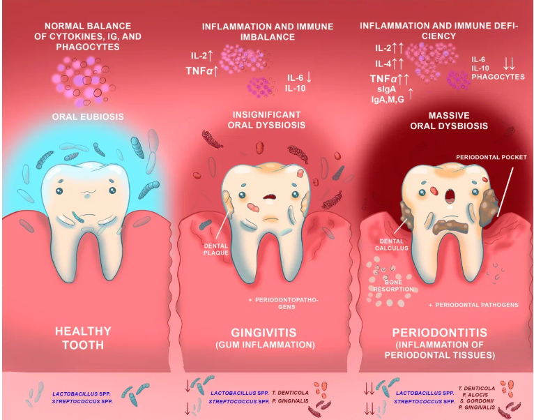
The data obtained indicate that the progression of IPD is associated with changes in the species composition of the oral microbiome, manifested in the predominance of periodontopathogen colonization in this biotope against the background of its reduced colonization by lactobacilli and streptococci. This fits into the logic of IPD pathogenesis, since it is known that lactobacilli, possessing high adhesive capacity and pronounced antagonistic activity, can compete with periodontopathogenic bacteria for attachment receptors on epithelial cells, inhibit their biofilm formation, and prevent their proliferation [28—31]. Evidently, the suppression of Lactobacillus spp. proliferation (and possibly that of some bacterial species of the genus Streptococcus) is associated with a decrease in the colonization resistance of the oral biotope, creating favorable conditions for the growth and multiplication of periodontopathogens of various species, thereby contributing to the development and progression of IPD with the involvement of the body’s immune system in the pathological process.
In this regard, we conducted an analysis of a number of immunological parameters in the examined individuals, which showed that in patients with IPD (gingivitis and CGP), the levels of pro-inflammatory (IL-2 and TNFα) and anti-inflammatory cytokines (IL-4), as well as sIgA, were elevated against the background of decreased levels of IL-10 and IL-6 and reduced phagocytic activity parameters (Table 2).
Table 2
Immunological indicators in the examined individuals
in the analyzed groups, Ме (Q1 and Q2)
|
Indicator |
Immunological indicators in the examined individuals |
|||
|
Healthy (n = 21) |
Gingivitis (n = 15) |
Mild CGP (n = 30) |
Moderate CGP (n = 39) |
|
|
IL-2, pg/mL |
11.3 (11.0; 11.6) |
12.8 (12.5; 13.1)* |
15.4 (14.3; 16.2)* |
26.5 (24.3; 27.0)* |
|
IL-4, pg/mL |
3.3 (3.1; 4.0) |
4.3 (4.3; 4.8) |
4.6 (4.2; 4.9) |
6.8 (6.1; 6.9)* |
|
IL-6, pg/mL |
1.0 (0.9; 1.0) |
0.9 (0.9; 1.0)* |
0.9 (0.7; 1.0)* |
0.8 (0.7; 0.8)* |
|
IL-10, pg/mL |
9.5 (8.9; 9.8) |
8.1 (7.8; 8.2)* |
4.1 (3.9; 4.5)* |
2.2 (2.0; 2.4)* |
|
TNFα, pg/mL |
3.3 (2.7; 4.2) |
9.5 (8.5; 15.6)* |
19.4 (17.5; 23.2)* |
44.8 (41.6; 48.8)* |
|
sIgA, g/L |
211.0 (159.8; 262.2) |
176.0 (138.9; 213.1) |
389.9 (327.7; 461.1)* |
466.4 (393.3; 539.5)* |
|
IgA, g/L |
1.6 (1.4; 1.9) |
1.8 (1.5; 2.3) |
2.0 (1.5; 2.3)* |
4.21 (3.7; 5.0)* |
|
IgМ, g/L |
1.4 (1.1; 1.6) |
1.4 (1.0; 1.6) |
1.3 (1.0; 1.6) |
2.1 (1.1; 3.6)* |
|
IgG, g/L |
13.5 (12.0; 15.0) |
12.9 (12.0; 13.8) |
14. 0 (9.6; 14.4) |
20.2 (15.8; 24.6)* |
|
Phagocytic index, % |
58 (56; 68) |
56 (52; 68) |
32 (27; 36)* |
29 (26; 36)* |
|
Phagocytic number, particles / cell |
8 (7; 9) |
9 (8; 9.5) |
3 (3; 4)* |
2.5 (2; 3)* |
Note: significant changes compared to the group of healthy individuals.
The levels of IL-2, IL-4, IL-6, IL-10, and TNFα were significantly dependent on the presence of a particular form of IPD in patients (χ² = 64.2…88.2, p << 0.0001), with the dynamics of IL-6 and IL-10 showing a ‘mirror’ pattern relative to the changes in IL-2, IL-4, and TNFα. The assessment of sIgA levels in the gingival sulcus / periodontal pocket content indicated a modification of the normal distribution of this parameter (Dmax = 0.9, p > 0.20); therefore, the analysis of the dependence of sIgA levels on the group affiliation of patients was performed using parametric Fisher’s analysis of variance. The dependence proved to be quite pronounced — η² = 75 %, F = 97.5 (p << 0.0001).
In addition, patients with CGP, compared to healthy individuals and patients with gingivitis, exhibited altered functional activity of peripheral blood leukocytes (Table 2), and the degree of reduction in the phagocytic index and phagocytic number depended on the group affiliation of the examined individuals (χ² = 55.0 and χ² = 68.3, respectively; p << 0.0001).
The modifications of the cytokine profile and sIgA levels revealed in patients with CGP apparently reflect manifestations of a mixed inflammatory response with a predominance of the pro-inflammatory reaction. The active nature of inflammation is indicated by the increased levels of pro-inflammatory cytokines (IL-2 and TNFα), which were simultaneously combined with an increase in the levels of the anti-inflammatory cytokine IL-4 and sIgA, which may reflect a compensatory mechanism aimed at regulating the inflammatory response. At the same time, the decreased levels of IL-10 (an important anti-inflammatory cytokine) and IL-6 (which has variable functional activity) probably confirm the insufficiency of anti-inflammatory control and indicate an imbalance in the immune response with a shift toward inflammation. Overall, the characterized deviations of these immunological parameters appear to indicate ineffective suppression of an already established and indolent inflammatory process in CGP.
Noteworthy is the fact that the transition of CGP from mild to moderate severity was accompanied by an increase in systemic humoral immunity parameters, in particular, the levels of serum immunoglobulins A, M, and G (Table 2). The serum levels of IgA and IgM in peripheral blood depended on the presence of IPD in patients — χ2 = 75.6 and χ2 = 48.6, respectively (p << 0.0001). The serum IgG level also depended on the group affiliation of patients (η2 = 40 %, F = 22.6, p << 0.0001), with a substantial and significant surge in the mean IgG level to 20.2 ± 4.4 g/L (p < 0.0001), as in the cases discussed above, occurring only in patients with moderate CGP.
Evaluating the totality of the presented data on the immunological parameters of the examined individuals (healthy and those with inflammatory periodontal pathology), it should be noted that the observed shifts in the immune status in patients with CGP apparently reflect a disruption of the balance between innate and adaptive immunity with a tendency toward immune response dysregulation and indicate the development of secondary immunodeficiency in patients with this pathology against the background of a torpid inflammatory course with prolonged antigenic stimulation by periodontopathogenic microorganisms.
This assumption was indirectly confirmed by the results of the correlation analysis, which revealed significant relationships between the occurrence of certain bacterial species in the oral cavity, as well as their associations, and the level of expression of individual immunological parameters.
Thus, the presence of V. dispar bacteria in the oral microbiome was accompanied by increased levels of TNFα, IL-2, IL-4, and sIgA (τ = 0.32, τ = 0.27, τ = 0.36, τ = 0.36, respectively; p < 0.05) and was also associated with a decrease in the phagocytic activity of neutrophils (τ = – 0.78, p < 0.01). The analysis of relationships between immunological parameters and bacteria of other species occurring in isolation in the gingival sulcus / periodontal pocket content revealed no significant correlations.
At the same time, it was established that the values of a number of immunological parameters correlate with the colonization of the oral cavity by two- and three-component associations of certain microorganisms. In particular, a negative correlation was revealed between the presence of F. nucleatum, F. alocis, and T. forsythia (in various combinations) in the oral biotope and the levels of TNFα, IL-2, IL-4, and sIgA (τ = – 0.57…– 0.86, p < 0.01), as well as humoral immunity parameters, namely IgA, IgM, and IgG (τ = – 0.67, τ = – 0.71, and τ = – 0.57, respectively; p < 0.01). It is possible that periodontopathogens may secrete substances that suppress cytokine production and sIgA synthesis, their sorption, and/or destruction (enzymatic cleavage by proteases), thereby subverting the immune response, which contributes to the persistence of the pathogens in the body and accounts for the chronic course and progression of IPD.
At the same time, a negative correlation was observed between the presence of L. fermentum / plantarum / reuteri / salivarius in the oral microbiome and immunological parameters such as IL-2, IL-4, TNFα, and sIgA (τ = – 0.34, τ = – 0.28, τ = – 0.36, τ = – 0.32, respectively; p < 0.05), and a positive correlation was observed with the phagocytic activity of leukocytes (τ = 0.78, p < 0.01). This may be associated with the ability of Lactobacillus spp. to inhibit pathogenic microorganisms by triggering an immune response [29—31] and/or to exhibit antagonistic activity against periodontopathogens.
Undoubtedly, further studies are required to confirm the identified patterns and to elucidate the mechanisms underlying the observed correlations.
Conclusion
The results of the study indicated substantial taxonomic changes in the oral microbiota in patients with gingivitis and chronic generalized periodontitis compared to healthy individuals, evidencing the development of pronounced dysbiotic disturbances in the oral cavity in inflammatory periodontal diseases. Not only individual microbial species were identified but also their associations, which likely play a decisive role in the etiology and pathogenesis of inflammatory periodontal diseases. The obtained data confirm the necessity of conducting metagenomic studies to investigate the structure of the oral microbiome, to identify novel periodontopathogens and/or etiologically significant multispecies microbial consortia, as well as to analyze the nature of their interactions (antagonism/synergism) and to assess the influence of the oral microbiota on the host immune response and its contribution to the development of concomitant (systemic) diseases, taking into account the changes in the immunological status of patients observed in inflammatory periodontal diseases, which affect both the humoral and cellular components of immunity and potentially facilitate the spread (translocation) of infection from the oral biotope into the body. The established level of genomic diversity and the identified distinctive characteristics of the oral microbiota may serve as a valuable tool for the diagnosis and prognosis of the course of inflammatory periodontal diseases, as well as for the development of personalized strategies and the enhancement of the efficacy of therapy and prevention of this pathology.
Reference
- Tarasenko S. V., Kulaga O. I., Vavilova T. P. Clinical and biochemical parallels in study of effectiveness of dental hyalrepair®-02 preparation in patients with chronic periodontitis. Medicinskij alfavit. 2016, 3(21), 5—10, EDN: WNDHIV. (in Russian).
- Gimranova I. A., Khakimova L. R., Akmalova G. M., Gazizullina G. R. Modern methods of diagnosis of periodontal diseases: opportunities and prospects (review of literature). Russian Clinical Laboratory Diagnostics. 2023, 68(9), 570—577, DOI: 10.51620/0869-2084-2023-68-9-570-577. (in Russian).
- Sharifovich A. D., Alimjanovich R. J. The role of the immune system and oral fluid and blood antimicrobial peptides in patients with chronic generalized periodontitis. The American Journal of Interdisciplinary Innovations and Research. 2023, 5, 64—71, DOI: 10.37547/TAJMSPR/Volume05Issue05-13.
- Cekici A., Kantarci A., Hasturk H., Van Dyke T. E. Inflammatory and immune pathways in the pathogenesis of periodontal disease. Periodontology. 2000, 2014, 64(1), 57—80, DOI: 10.1111/prd.12002.
- Hajishengallis G. Immunomicrobial pathogenesis of periodontitis: keystones, pathobionts, and host response. Trends in immunology. 2014, 35(1), 3—11, DOI: 10.1016/j.it.2013.09.001.
- Devine D. A., Marsh P. D., Meade J. Modulation of host responses by oral commensal bacteria. J. Oral. Microbiol. 2015, 7, 26941, DOI: 10.3402/jom.v7.26941.
- Wang R., Hu J., Sun Q., Guo S., Zhang G., Lu A., et al. Identification and validation of endoplasmic reticulum autophagy-related potential biomarkers in periodontitis. Sci. Rep. 2025, 15(1), 24151, DOI: 10.1038/s41598-025-08180-2.
- Faisal R. H., Ali A. O. Increased bacterial load of Filifactor alocis in deep periodontal pockets discriminate between periodontitis stage 3 and 4. Front. Oral Health. 2025, 6, 1543030, DOI: 10.3389/froh.2025.1543030.
- Tsarev V. N., Arutunov S. D., Balmasova I. P., Babaev E. A., Nikolaeva E. N., Ippolitov E. V., et al. Molecular diagnostic of periodontitis and metagenomic analysis of the periodotum microbiota in patients by type II diabetes mellitus. Bacteriology. 2018, 3(2), 30—37, DOI: 10.20953/2500-1027-2018-2-30-37. (in Russian).
- Tsarev V. N., Nikolaeva E. N., Ippolitov E. V. Periodontophatogenic bacteria of the main factors of emergence and development of periodontitis. Journal of Microbiology, Epidemiology and Immunobiology. 2017, 5, 101—112, DOI: 10.36233/0372-9311-2017-5-101-112. (in Russian).
- Chuykin S. V., Mavzyutov A. R., Chuykin O. S., Akat’yeva G. G., Kuchuk K. N. Study of periodontal pathogens by polymerase chain reaction in children with congenital cleft palate and a postoperative defect. Pediatric dentistry and dental prophylaxis. 2022, 22(1), 19—28, DOI: 10.33925/1683-3031-2021-22-1-19-28. (in Russian).
- Bulgakova А. I., Mavzyutov A. R., Tamarova E. R., Vasiljeva N. A., Shvetz К. Yu. Сlinical and microbiological substantiation of a complex treatment of patients with periodontitis with moderate and severe severity in view of molecular genetic characteristics oral microbiota. Periodontology (Russia). 2017, 22(1), 70—73, EDN: YPSSWH (in Russian).
- Tsarev V. N., Atrushkevich V. G., Ippolitov E. V., Podporin M. S. Comparative analysis of periodontal antiseptic antimicrobial activity using the automated control of microbial growth in real-time. Periodontology (Russia). 2017, 22(1), 4—10, EDN: YPSSQN. (in Russian).
- Domenyuk D. A., Davydov B. N., Gilmiyarova F. N., Ivchenko L. G. The influence of severity of diabetes mellitus type i in children on the dental status and the immunological, biochemical indicators of blood serum and oral fluid. Part IPeriodontology (Russia). 2017, 2(83), 53—60, EDN: YPRZSP (in Russian).
- Dzyuba E., Nagaeva M., Zhdanova E. The role of immunological processes in the development of inflammatory diseases of periodontal disease and the possibilities of their correction. Actual problems in dentistry. 2019, 15(2), 24—31, DOI: 10.18481/2077-7566-2019-15-2-25-31. (in Russian).
- Pan W., Wang Q., Chen Q. The cytokine network involved in the host immune response to periodontitis. Int. J. Oral Sci. 2019, 11(3), 30, DOI: 10.1038/s41368-019-0064-z.
- Plemmenos G., Evangeliou E., Polizogopoulos N., Chalazias A., Deligianni M., Piperi C. Central regulatory role of cytokines in periodontitis and targeting options. Curr. Med. Chem. 2021, 28(15), 3032—3058, DOI: 10.2174/0929867327666200824112732.
- Bykova N. I., Odolski A. V., Grigoryan V. A. Indicators of cellular and humoral immunity in experimental inflammation of periodontal tissues. Kuban scientific medical bulletin. 2016, 6, 20—26, EDN: XVJHCH. (in Russian).
- Tsybikov N. N., Domanova E. T., Zobnin V. V., Ignatov M. Yu., Maslo E. Yu., Isakova N. V. The properties of the gingival fluid from the patients presenting with acute gingivitis and chronic periodontitis. Russian journal of dentistry. 2012, 1, 40—42, EDN: PGJEVJ. (in Russian).
- Linhartova P. B., Danek Z., Deissova T., Hromcik F., Lipovy B., Szaraz D., et al. Interleukin gene variability and periodontal bacteria in patients with generalized aggressive form of periodontitis. Int. J. Mol. Sci. 2020, 21(13), 4728, DOI: 10.3390/ijms21134728.
- Ovcharenko E. S., Erichev V. V., Aksyonova T. V., Melekhov S. V. Immunological and microbiological evaluation of oral hygiene effectiveness in complex treatment of patients with inflammatory periodontal diseases. Medicinskij alfavit. 2017, 3(24), 38—41, EDN: ZVRCZT. (in Russian).
- Callahan B. J., McMurdie P. J., Rosen M. J., Han A. W., Johnson A. J. A., Holmes S. P. DADA2: High-resolution sample inference from Illumina amplicon data. Nat. Methods. 2016, 13, 581—583, DOI: 10.1038/nmeth.3869.
- Quast C., Pruesse E., Yilmaz P., Gerken J., Schweer T., Yarza P., et al. The SILVA ribosomal RNA gene database project: Improved data processing and web-based tools. Nucleic Acids Res. 2013, 41(Database issue), D590—D596, DOI: 10.1093/nar/gks1219.
- Grjibovski A. M. Analysis of nominal data (Independent observations). Human Ecology. 2008, 8, 58—68, EDN: KXIIFZ. (in Russian).
- Benahmed A. G., Mujawdiya P. K., Noor S., Gasmi A. Porphyromonas gingivalis in the development of periodontitis: impact on dysbiosis and inflammation. Arch. Razi Inst. 2022, 77(5), 1539—1551, DOI: 10.22092/ARI.2021.356596.1875.
- Hajishengallis G., Liang S., Payne M. A., Hashim A., Jotwani R., Eskan M. A., et al. Low-abundance biofilm species orchestrates inflammatory periodontal disease through the commensal microbiota and complement. Cell Host Microbe. 2011, 10(5), 497—506, DOI: 10.1016/j.chom.2011.10.006.
- Bazhutova I. V., Ismatullin D. D., Lyamin A. V., Trunin D. A., Zhestkov A. V., Razumnyj V. A. Clinical significance of STREPTOCOCCUS members in developing periodontitis. Russian Journal of Infection and Immunity. 2022, 12(1), 51—58, DOI: 10.15789/2220-7619-CSO-1698. (in Russian).
- Jamal M., Ahmad W., Andleeb S., Jalil F., Imran M., Nawaz M. A., et al. Bacterial biofilm and associated infections. J. Chin. Med. Assoc. 2018, 81(1), 7—11, DOI: 10.1016/j.jcma.2017.07.012.
- Barzegari A., Kheyrolahzadeh K., Khatibi S. M.H., Sharifi S., Memar M. Y., Vahed S. Z. The battle of probiotics and their derivatives against biofilms. Infect. Drug. Resistance. 2020, 13, 659—672, DOI: 10.2147/idr.s232982.
- Gou H.-Z., Zhang Y.-L., Ren L.-F., Li Z.-J., Zhang L. How do intestinal probiotics restore the intestinal barrier? Front. Microbiol. 2022, 13, 929346, DOI: 10.3389/fmicb.2022.929346.
- Vasiee A., Falah F., Mortazavi S. A. Evaluation of probiotic potential of autochthonous lactobacilli strains isolated from Zabuli yellow kashk, an Iranian dairy product. J. Appl. Microbiol. 2022; 133(5), 3201—3214, DOI: 10.1111/jam.15772.