
The role of Fcγ-receptors in the biological action of C-reactive protein on human mast cells
- DOI
- 10.5922/ATB-2026-2-1-2
- Pages
- 17-45
- To cite
- Trulioff A. S., Kutukova N. A., Kudriavtsev I. V. The role of Fcγ-receptors in the biological action of C-reactive protein on human mast cells. Advanced Targets in Biomedicine. 2026;2(1):17–45.
Abstract
Full text
Introduction
Mast cells (MCs) are multifunctional immune cells involved in allergic reactions as well as in a wide range of physiological and pathological processes. These cells emerged early in evolution: their counterparts have been identified in ascidians and arthropods, where they perform functions similar to those of mammalian mast cells [1]. Recent studies have expanded our understanding of the role of mast cells, confirming their involvement in antiviral and antibacterial responses, the induction of immune tolerance, angiogenesis, and tissue repair. Mast cell dysfunction has been associated with the development of rheumatoid arthritis [2], multiple sclerosis [3], and fibrotic diseases [5].
Mast cell localization in the body follows a distinct pattern. They are concentrated near epithelial barriers such as the skin and the mucosa of the gastrointestinal and respiratory tracts. Their distribution follows two general rules: density increases closer to the epidermis and in distal areas (facial skin, palms), which correlates with an increased risk of pathogen contact [6]. Unlike other hematopoietic cells, mature mast cells do not circulate in the blood but reside permanently within tissues. The mechanisms underlying their migration remain a matter of debate, although interactions between adhesion molecules and extracellular matrix components are thought to play a key role in this process [7].
Fibroblasts, the principal stromal cells of connective tissue, create the microenvironment required for mast cell maturation and function. Mast cell activation initiates a cascade of inflammatory reactions: the release of mediators (histamine, cytokines) increases vascular permeability, which clinically manifests as the classical tetrad of inflammation: redness, swelling, heat, and pain [8].
Of particular interest are the pentraxins, acute-phase proteins that include C-reactive protein (CRP) and serum amyloid P component (SAP). These oligomeric structures perform an opsonizing function, bind apoptotic cells, and regulate complement activity [9]. The dynamics of their synthesis during inflammation differ: CRP levels increase 100- to 1000-fold (up to 200 μg/ml), whereas SAP concentrations fluctuate only slightly (around 30 μg/ml) [10]. CRP interacts with a broad range of ligands, including fibronectin, laminin, and pathogen-associated molecules, indicating its role in modulating intercellular interactions [11].
Despite extensive data on the pro-inflammatory activity of mast cells and the reparative function of fibroblasts, their cooperation during the early stages of inflammation remains poorly understood. The receptor mechanisms mediating the effects of CRP on connective tissue cells are still a matter of debate.
Aim of the study
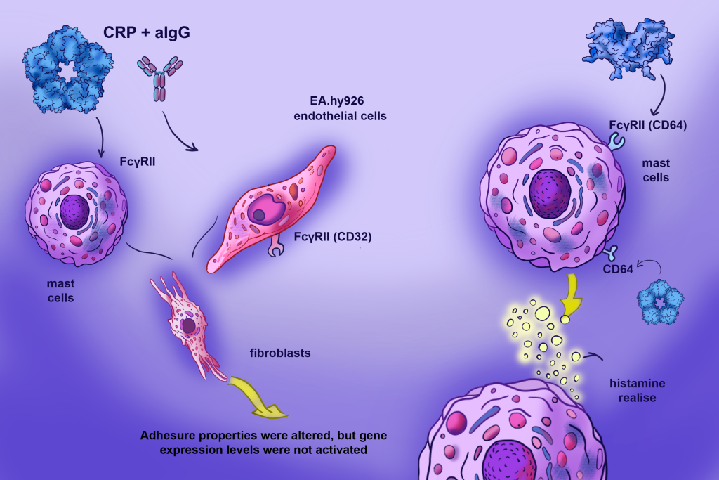
The study aimed to investigate the role of CRP in regulating the interactions of mast cells with fibroblasts and endothelium, and to assess the involvement of Fcγ receptors in these processes.
Materials and methods
Cell cultures
The HMC-1 mast cell line was kindly provided by Prof. J. H. Butterfield (Mayo Clinic, Rochester, USA). Cells were cultured in Iscove’s Modified Dulbecco’s Medium (IMDM, Biolot, St. Petersburg) supplemented with 10 % fetal calf serum (FCS), gentamicin (40 μg/ ml), and 1.2 mM monothioglycerol. Cells were subcultured every 3—4 days upon reaching a density of 1.5—2 × 10⁶ cells/ml.
Human fibroblasts were kindly provided by D. A. Mogilenko. Fibroblasts were isolated from the foreskin of healthy donors. EA.hy926, an immortalized human endothelial cell line, was kindly provided by Dr. C.-J. S. Edgell (University of Northern California, USA ). Both EA.hy926 endothelial cells and fibroblasts are adherent cultures. Cells were cultured in DMEM (Sigma) supplemented with 10 % fetal calf serum (Gibco), 40 μg/ml gentamicin sulfate (Biolot, St. Petersburg, Russia), and 2 mM L-glutamine (Biolot, St. Petersburg, Russia). Cells were subcultured upon reaching a confluent monolayer, 2—3 times per week. A 1 : 1 trypsin—Versene solution (Biolot, St. Petersburg, Russia) was used to disaggregate the monolayer.
All cell cultures used in this study were incubated in 50-ml polystyrene flasks (Orange Scientific, Belgium) at 37 °C in a 5 % CO2 atmosphere.
Assessment of the cytotoxic effect of the test preparations on HMC-1 cells
CRP (Calbiochem) and IgG aggregated by heating at 63 °C (ImTek) were used in this study. The potential effect of the drug doses used in the experiments on the viability of HMC-1 cells was tested. Cytotoxicity was assessed by flow cytometry, based on the appearance of cells with hypodiploid DNA content following propidium iodide staining. CRP and aIgG were tested at final concentrations of 5, 10, and 50 μg/ml.
After incubation with the test substances, cells were washed with excess phosphate-buffered saline (PBS). The resulting pellet was resuspended in 100 μl of PBS, and 900 μl of 70 % ethanol, pre-cooled to – 20 °C, was then added. Immediately before analysis, cells were pelleted by centrifugation (7 min at 300 g) and resuspended in PBS. A 0.1 % propidium iodide solution was then added to the samples, which were stained for 30 min in the dark at room temperature. After incubation, samples were analyzed by flow cytometry using a Navios flow cytometer (Beckman Coulter Inc., USA).
Flow cytometry
Expression of various surface molecules was assessed using a Navios flow cytometer (Beckman Coulter) with monoclonal antibodies. Corresponding isotype controls were used for each monoclonal antibody (CD16, CD32, CD64, CD11b, and CD54). All antibodies and isotype controls used in this study were manufactured by Beckman Coulter Inc. (USA). Staining of surface antigens with antibodies was performed according to the manufacturer’s instructions. Results were expressed as arbitrary units of MFI (geometric mean), reflecting mean fluorescence intensity.
Assessment of mast cell activation by histamine release (Shore method)
To assess the ability of Fcγ receptor ligands to induce mast cell degranulation, the required number of cells was collected, washed with Hanks’ Balanced Salt Solution (HBSS, Biolot), and resuspended in HBSS containing 0.1 % bovine serum albumin (BSA, Sigma). The substances of interest were then added to the cells, and compound 48/80 (Sigma) at a concentration of 20 μg/ml was used as a positive control. After a 30-minute incubation at 37 °C, cells were pelleted by centrifugation (5 min at 300 g), and the supernatant was collected and frozen at – 20 °C. To determine total cellular histamine content, cells were diluted with deionized water and disrupted by freeze—thaw cycles.
The amount of released histamine was determined by a modified Shore method. The result was expressed as the ratio of the RFU in the supernatant to the sum of the RFU in the supernatant and the RFU of histamine remaining in the cells.
Assessment of Cell—cell adhesion
Adhesion of HMC-1 cells to adherent cultures was assessed as follows: CFSE-pre-labeled HMC-1 cells were added to the wells of a 96-well plate containing a monolayer of adherent cells. CFSE-labeled suspension cells were co-incubated with a monolayer of fibroblasts (Fb) or endothelial cells for 30 min, after which the monolayer was washed to remove unattached cells and the reaction was quantified.
When studying the effect of Fcγ receptor ligands on the adhesive properties of the suspension cells, cells were stimulated with the test agent for 24 hours and then washed and labeled with CFSE.
When studying stimulator-induced changes in the adhesive properties of fibroblasts or EA.hy926 cells, inducers diluted in serum-free medium containing 1 % BSA were added 24 hours before the introduction of CFSE-labeled cells. Adhesion was quantified using a spectrofluorometer at an excitation wavelength of 480 nm and an emission wavelength of 535 nm. The number of attached cells — i. e., the degree of adhesion — was estimated from the fluorescence intensity. Results were expressed in RFU.
When studying cell—cell adhesion in the presence of inducers, CFSE-labeled cells were added to wells containing adherent cells in 50 μl of culture medium, immediately followed by the addition of stimulators in 50 μl of the same medium.
Binding of C-reactive protein to cells Biotin labeling of C-reactive protein
To label CRP with biotin, the commercial CRP preparation was dialyzed twice against PBS. A 1 % biotin solution (sulfosuccinimidyl-6-[biotin]hexanoate [Sulfo-NHS-LS-Biotin], Pierce) in dH2O was then added to CRP at a molar ratio of CRP to biotin of 1:20. After a 30-minute incubation at room temperature, the labeled CRP was dialyzed three times against PBS to remove unbound biotin; the final dialysis step included 0.2 % sodium azide (Serva).
Binding of C-reactive protein to HMC-1 mast cells
CRP binding to HMC-1 mast cells was assessed by flow cytometry. Cells were collected and washed with HBSS containing 0.1 % BSA and 0.2 % NaN3. Biotin-labeled CRP was then added to the cells at various concentrations and incubated for 30 min at room temperature. Cells were then washed twice to remove unbound CRP, and 0.5 μl of allophycocyanin (APC)-conjugated streptavidin was added; samples were incubated in the dark at room temperature for 15 min. After incubation, cells were washed three times with PBS containing 10 % fetal calf serum, and fluorescence intensity was measured by flow cytometry.
Competition experiments were also performed to examine competition between biotinylated and unlabeled CRP or aIgG for cell binding. In these experiments, 50 μg/ml biotin-labeled CRP and various concentrations of unlabeled CRP or aIgG were added to cells simultaneously, followed by a 30-minute incubation, washing, addition of APC-conjugated streptavidin, and measurement of cell fluorescence by flow cytometry. The concentration of biotin-labeled CRP was chosen because, at this concentration, CRP binding to cells was clearly distinguishable from the control but had not yet reached saturation (Fig. 1, a).
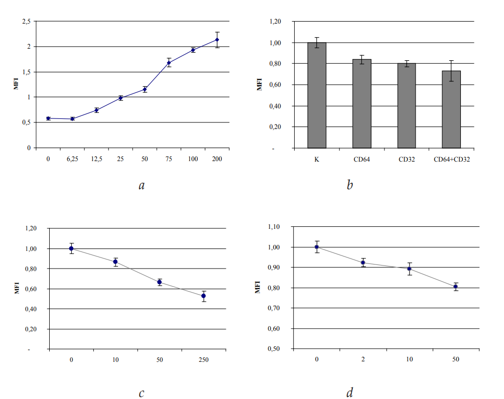
Fig. 1. Binding of C-reactive protein to HMC-1 cells: a — CRP binding to HMC-1 cells;
abscissa — CRP concentration, μg/ml; ordinate — fluorescence intensity
of cells stained with streptavidin-APC, MFI;
b — Evaluation of the specificity of CRP binding to HMC-1 mast cells: displacement
of biotin-labeled CRP by antibodies against FcγR; ordinate — MFI;
c — Displacement of biotin-labeled CRP by increasing concentrations of unlabeled CRP;
abscissa — concentration of unlabeled CRP, μg/ml; ordinate — MFI;
d —Evaluation of the specificity of CRP binding to HMC-1 mast cells:
displacement of biotin-labeled CRP by increasing concentrations of aIgG;
abscissa — concentration of aIgG, μg/ml; ordinate — MFI
Transfection
To assess activation of the NF-κB signaling pathway, HMC-1 mast cells were transfected with the pNF-κBluc and pCMVL plasmids using Lipofectamine 2000 (Invitrogen) according to the manufacturer’s instructions. The pNF-κBluc plasmid contained the luciferase gene (luc) linked to a minimal synthetic promoter containing five NF-κB cis-elements (Stratagene, USA). The pCMVL plasmid contained the β-galactosidase gene under the control of the human cytomegalovirus (CMV) promoter. Plasmids were isolated from E. coli cells by alkaline lysis. Twenty-four hours after transfection, cells were incubated for 24 hours with human CRP (Calbiochem/MD Biochemicals), human serum amyloid P component (SAP) (Calbiochem), or aggregated human IgG (ImTek, Moscow). After incubation with the stimulators, cells were washed three times with PBS, harvested, and lysed in 20 μl of lysis buffer (Promega).
Statistical analysis
Statistical analysis of normally distributed data was performed using MS Excel 2010 (Microsoft, USA). The normality of the data distribution was assessed using the Shapiro—Wilk and Kolmogorov—Smirnov tests. Results are presented as the arithmetic mean and its standard error (M±m). The significance of differences between groups was assessed by one-way ANOVA (Welch’s ANOVA). Dunnett’s post hoc test (for comparisons with the control group) and Tamhane’s T2 test (for all pairwise comparisons) were used for multiple comparisons. Differences were considered statistically significant at p < 0.05.
Results
1. Toxic effect of different doses of C-reactive protein and aggregated human IgG on HMC-1 cells
Experiments were performed to assess the potential cytotoxicity of C-reactive protein (CRP) and aggregated human IgG antibodies (aIgG) at concentrations ranging from 5 to 50 μg/ ml. Bovine serum albumin at a concentration of 100 μg/ml was used as a negative control. As shown in Table 1, 24-hour exposure to the studied Fcγ receptor ligands did not result in a statistically significant increase in apoptotic activity of HMC-1 cells. This indicates the absence of a direct toxic effect of CRP and aIgG on cell viability at the selected concentrations. Consequently, the effects observed in subsequent experiments cannot be attributed to non-specific cell death by necrosis or apoptosis.
Table 1
Toxic effect of different doses of CRP and aIgG on HMC-1 cells
Stimulator | Cells with hypodiploid DNA content, % |
Control: BSA, 100 ng/ml | 7.9 ± 1.5 |
CRP, 5 μg/ml | 6.2 ± 2.1 |
CRP, 10 μg/ml | 6.9 ± 3.5 |
CRP, 50 μg/ml | 7.0 ± 1.2 |
aIgG, 5 μg/ml | 7.7 ± 1.4 |
aIgG, 10 μg/ml | 6.3 ± 3.6 |
aIgG, 50 μg/ml | 8.3 ± 2.6 |
Note: Data are presented as M±m; n=6.
2. Expression of Fcγ receptors on the studied cell lines
Surface markers were analyzed by flow cytometry. HMC-1 cells were found to show pronounced expression of CD32 (FcγRII). Moderate levels of CD64 (FcγRI) were detected on their surface, whereas CD16 (FcγRIII) was absent (Fig. 2).
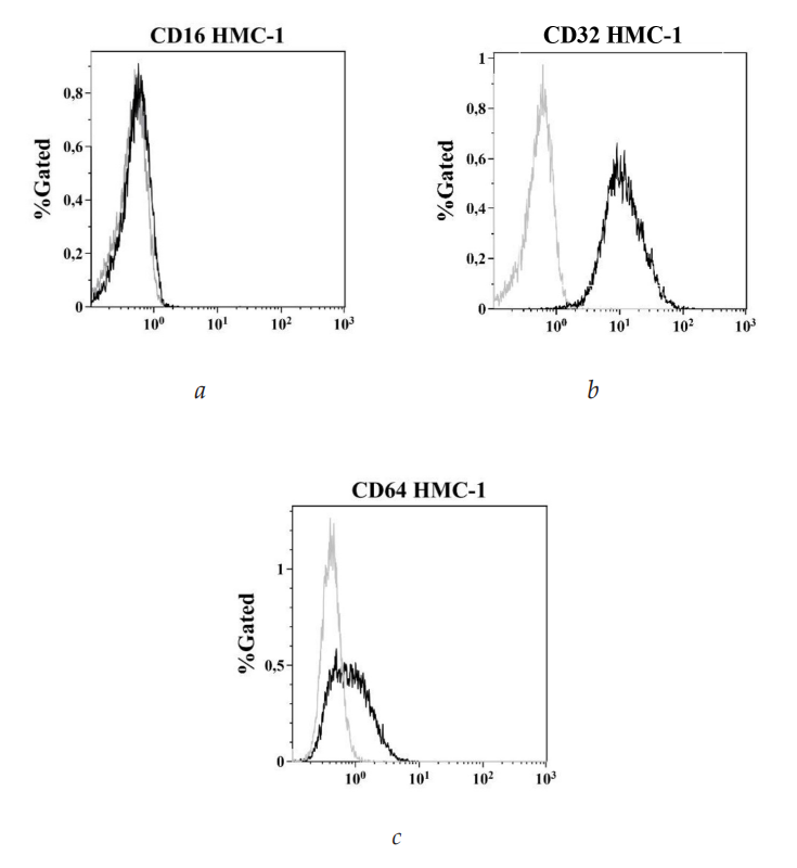
Fig. 2. Expression of Fcγ receptors by HMC-1 cells : a — Binding of HMC-1 cells to anti-CD16 antibodies (FcγRIII); b — Binding of HMC-1 cells to anti-CD32 antibodies (FcγRII);
c — Binding of HMC-1 cells to anti-CD64 antibodies (FcγRI)
Note: gray indicates the level of cell binding to isotype control antibodies; black indicates binding of antibodies against the corresponding Fcγ receptors. The abscissa shows fluorescence intensity (MFI); the ordinate shows cell number.
3. Effect of IFNγ on Fcγ receptor expression by HMC-1 cells
Incubation of HMC-1 cells with interferon-gamma (IFNγ) was found to induce an increase in expression of the high-affinity receptor CD64 (FcγRI). Maximum expression was observed 24 hours after stimulation (Fig. 3, a). The effect of IFNγ was dose-dependent: no changes were observed at 1 ng/ml, whereas 10 ng/ml increased CD64 expression by 20 %, and 100 ng/ml by 28 %. A further increase in cytokine dose (up to 1000 ng/ml) did not enhance the response, indicating that a plateau had been reached (Fig. 3, b).
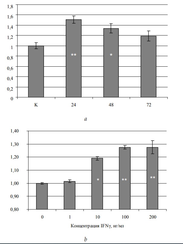
Fig. 3. FcγRI expression by HMC-1 mast cells in response to IFNγ: a — Effect of stimulation duration; abscissa — duration of cell incubation with IFNγ (15 ng/ml), hours; ordinate — fluorescence intensity of cells stained with FITC-conjugated anti-CD64 antibodies (MFI); b — Dependence of FcγRI expression on IFNγ concentration; abscissa — IFNγ concentration; ordinate — fluorescence intensity of cells stained with FITC-conjugated
anti-CD64 antibodies (MFI)
Note: significance of differences from control: * — p < 0.05, ** — p < 0.01, *** — p < 0.001.
IFNγ exposure had no significant effect on the expression of the low-affinity receptors CD32 (FcγRII) and CD16 (FcγRIII) (Fig. 4, a, b).
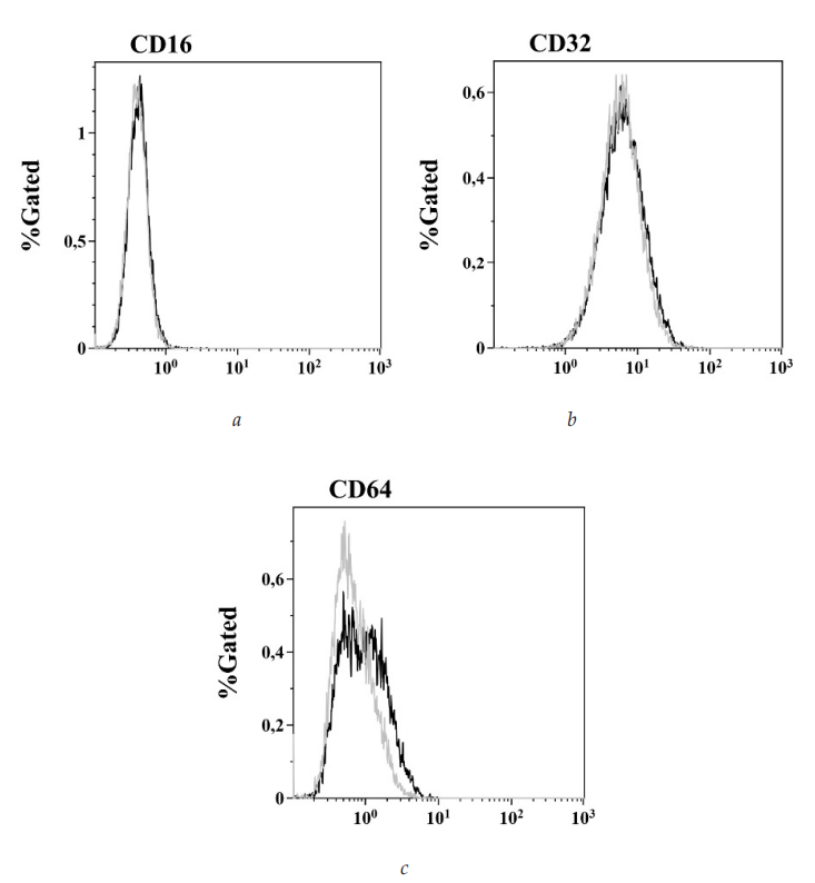
Fig. 4. Expression of Fcγ receptors by HMC-1 cells. Effect of IFNγ:
a — Expression of CD16 (FcγRIII) on HMC-1 cells;
b — Expression of CD32 (FcγRII) on HMC-1 cells;
c — Expression of CD64 (FcγRI) on HMC-1 cells. Binding
of antibodies against the corresponding
Fcγ receptors to intact cells is shown in gray; binding to IFNγ-activated cells is shown in black
Note: the abscissa shows fluorescence intensity (MFI); the ordinate shows cell number.
4. Binding of C-reactive protein to HMC-1 mast cells
Our experimental data demonstrated the ability of C-reactive protein to bind to HMC-1 mast cells (Fig. 5). This process did not require the presence of Ca2+ or Mg2+ ions in the medium, as shown by the results presented in Table 2.
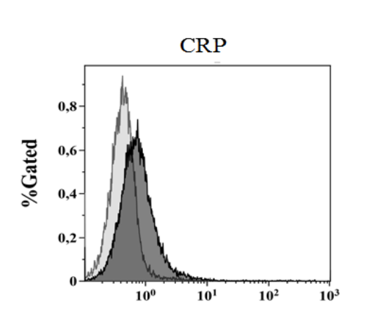
Fig. 5. Binding of C-reactive protein to HMC-1 cells. C-reactive protein was labeled with biotin and added to the cells at a concentration of 100 μg/ml; after a 30-minute incubation, unbound protein was washed off and APC-conjugated streptavidin was added (dark histogram); the light histogram shows streptavidin-APC added to cells without prior incubation with CRP (control)
Note: the abscissa shows fluorescence intensity (MFI); the ordinate shows cell number.
Table 2
CRP binding to HMC-1 mast cells:
independence from cation availability and IFNγ cell activation
Medium composition | Control | After IFNγ activation |
DPBS with Ca2+ and Mg2+ cations | 1.47 | 1.22 |
DPBS with 0.5 mM EDTA | 1.4 | 1.35 |
Note: Fluorescence intensity of cells that bound CRP is shown. Data are expressed in MFI. DPBS — Dulbecco’s phosphate-buffered saline. Differences from the control are not significant (p > 0.05).
The analysis showed no substantial effect of the medium’s cation composition or of IFNγ stimulation on the intensity of CRP binding to the cell surface (Table 2). Interaction was detected using a streptavidin-APC conjugate followed by flow cytometric analysis. The threshold CRP concentration at which specific binding was detected was 12.5 μg/ml. In the range of 12.5—100 μg/ml, a linear relationship was observed between protein dose and binding intensity. Above 100 μg/ml, the process reached a plateau, and further increases in concentration did not produce a statistically significant rise in fluorescence intensity (see Fig. 1, a).
Blocking Fcγ receptors with antibodies produced a modulating effect. Incubation with anti-CD64 monoclonal antibodies reduced CRP binding by 16 %, whereas anti-CD32 antibodies reduced it by 20 %. Combined treatment with inhibitors of both receptors produced an additive effect, reducing binding by 27 % (see Fig. 1, b).
Competitive inhibition experiments confirmed the selectivity of C-reactive protein binding to HMC-1 mast cells. In a system where biotin-labeled CRP (50 μg/ml) competed with the native form of the protein, the addition of 10 μg/ml unmodified CRP reduced the fluorescent signal by 14 %. Increasing the competitor concentration to 50 μg/ml enhanced the inhibitory effect, reducing binding by 33 %. Maximal suppression of binding (48 %) was observed at a five-fold molar excess of unlabeled protein relative to the labeled form (see Fig. 1, c).
Further experiments revealed the effect of aggregated IgG (aIgG) on CRP binding to the surface of HMC-1 mast cells. Co-incubation of cells with aIgG and biotinylated CRP produced a dose-dependent decrease in fluorescence. At an aIgG concentration of 2 μg/ ml, binding was suppressed by 8 %, whereas 10 μg/ml aggregated immunoglobulin reduced it to 11 %. The maximal inhibitory effect (20 %) was recorded at 50 μg/ml aIgG, indicating a partially competitive interaction mechanism (see Fig. 1, d).
5. Effect of C-reactive protein on degranulation of HMC-1 mast cells
In the absence of prior stimulation, CRP at concentrations of 0.03—30 μg/ml did not induce statistically significant histamine release from HMC-1 mast cells. However, after pre-activation of cells with interferon-gamma (IFNγ), a pronounced dose-dependent modulation of degranulation was observed.
Specifically, CRP at concentrations of 0.03 and 0.3 μg/ml increased histamine secretion by 18 % and 20 %, respectively, relative to the control. Increasing the protein dose to 3 μg/ml raised the degranulation level by 33 %, and at a CRP concentration of 30 μg/ml, histamine secretion reached 123 % of control values (Fig. 6).
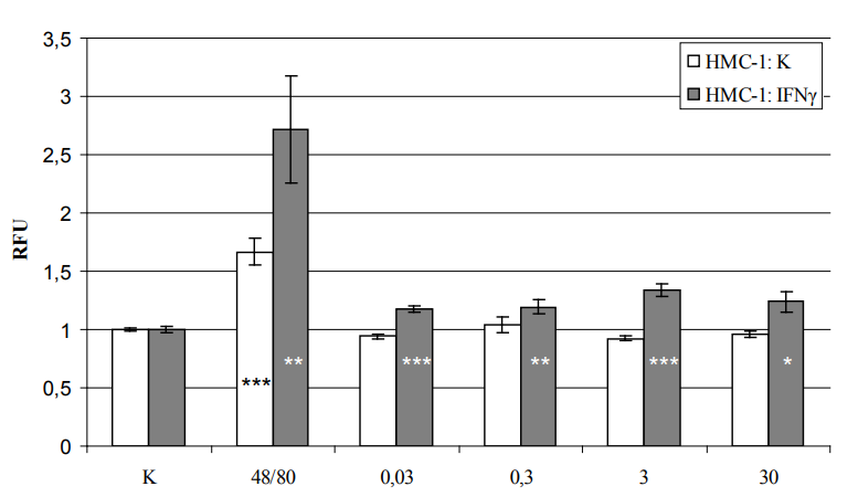
Fig. 6. Effect of CRP on histamine release from HMC-1 mast cells: dependence on IFNγ.
HMC-1 cells were incubated for 24 hours with 15 ng/ml IFNγ (gray bars) or without IFNγ
(white bars) and then stimulated with CRP at various concentrations. Abscissa:
C — PBS (negative control); 48/80 — compound 48/80 at 20 μg/ml (positive control); numbers — CRP concentration, μg/ml. Ordinate — stimulation index
(ratio of histamine release induced by the stimulator to the negative control).
The level of spontaneous histamine release was 0.220 ± 0.01
in the control without IFNγ treatment and 0.195 ± 0.02 after IFNγ treatment (p < 0.05)
Note: significance of differences from control: * — p < 0.05, ** — p < 0.01, *** — p < 0.001.
6. Effect of Fcγ receptor ligands (C-reactive protein, aggregated IgG) on cell adhesion
6.1. Effect of C-reactive protein and aggregated IgG on the adhesive properties
of HMC-1 mast cells
Experimental data demonstrated that 24-hour incubation of HMC-1 mast cells with C-reactive protein produced a statistically significant, CRP-concentration-dependent increase in their adhesive capacity. At a CRP dose of 10 μg/ml, MC adhesion to unstimulated fibroblasts increased by 27 % relative to control values. Increasing the protein concentration to 50 μg/ml raised this value by 54 %, with the differences between groups being statistically significant (p < 0.05) (Fig. 7).
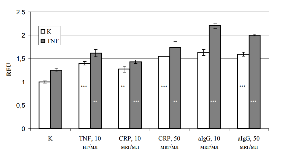
Fig. 7. Adhesion of stimulated HMC-1 mast cells to intact or TNF-activated fibroblasts
Note: white bars — adhesion of HMC-1 cells to intact fibroblasts; gray bars — adhesion to fibroblasts activated with TNF (10 ng/ml). Abscissa — mast cell activators and their concentrations; ordinate — stimulation index. Differences from control are significant: ** — p < 0.01; *** — p < 0.001.
Stimulation of MCs with aggregated IgG (aIgG) at concentrations of 10 and 50 μg/ ml also induced enhanced adhesion to fibroblasts (Fb). In both cases, the increase in adhesive activity was approximately 60 % compared with control.
To model the inflammatory process in tissues, cell cultures were stimulated separately. Fibroblasts were treated with TNF at 10 ng/ml, while MCs were incubated with CRP (10 or 50 μg/ml), aIgG (equivalent doses), or TNF (10 ng/ml). The results showed that both CRP and aIgG enhanced MC adhesion to fibroblasts, and that adhesion to TNF-activated Fb was significantly more pronounced than to intact cells. Relative to control, CRP at 10 μg/ ml increased adhesion by 14 %, and at 50 μg/ml by 38 %; aIgG at 10 and 50 μg/ml increased adhesion by 65 % and 49 %, respectively.
Comparison with the baseline level (intact MCs + intact Fb) showed that CRP stimulation at 10 μg/ml enhanced adhesion to TNF-activated Fb by 42 %, and at 50 μg/ml by 73 %. Treatment with aIgG (10 and 50 μg/ml) increased adhesion by 120 % and 100 %, respectively. Combined exposure of fibroblasts to TNF and of mast cells to FcγR ligands produced an additive effect, resulting in an additional 20 % increase in adhesion compared with intact Fb.
Based on the results of the preceding experiments, the effect of CRP and aIgG on the expression of the adhesion molecules CD11b (integrin) and CD54 (ICAM-1) on the surface of HMC-1 mast cells was analyzed. The results showed no significant changes in the expression of these markers upon incubation of cells with CRP (10—50 μg/ml) or aIgG (equivalent concentrations) for the times tested (Fig. 8, a, b, d, e). In contrast, TNF stimulation induced a statistically significant increase in CD54 expression on the membrane of HMC-1 MCs (Fig. 8, c).
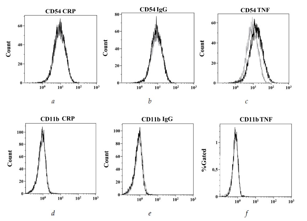
Fig. 8. Expression of CD54 and CD11b by HMC-1 cells stimulated with CRP, aIgG, and TNF:
a, b, c — ICAM-1 expression by HMC-1 cells stimulated with CRP (a), aIgG (b),
and TNF (c); d, e — CD11b expression by cells stimulated with CRP (d) or aIgG (e)
Note: gray indicates binding of antibodies against ICAM-1 or CD11b to intact cells; black indicates binding of antibodies to stimulated cells. The abscissa shows fluorescence intensity (RFU); the ordinate shows cell count.
Comparative analysis of the histograms (Fig. 8) confirmed that the CD54 expression profiles of the control group and of cells treated with CRP or aIgG were fully superimposable. In contrast, TNF activation of cells produced a marked shift in the histogram, indicating increased expression of this marker.
6.2. Effect of C-reactive protein and aggregated IgG on the adhesiveness
of fibroblasts for HMC-1 mast cells
It was experimentally established that pretreatment of fibroblasts (Fb) with CRP, despite fibroblasts lacking Fcγ receptors (see above), induced a dose-dependent increase in the adhesion of intact HMC-1 mast cells. At a CRP concentration of 5 μg/ ml, adhesion levels remained comparable to control, whereas increasing the dose to 10 and 50 μg/ml enhanced MC attachment by 11 % and 123 %, respectively (p < 0.05) (Fig. 9). In contrast to CRP, incubation of Fb with aggregated IgG (aIgG) at equivalent concentrations (10—50 μg/ml) did not produce statistically significant changes in adhesion (Fig. 10).
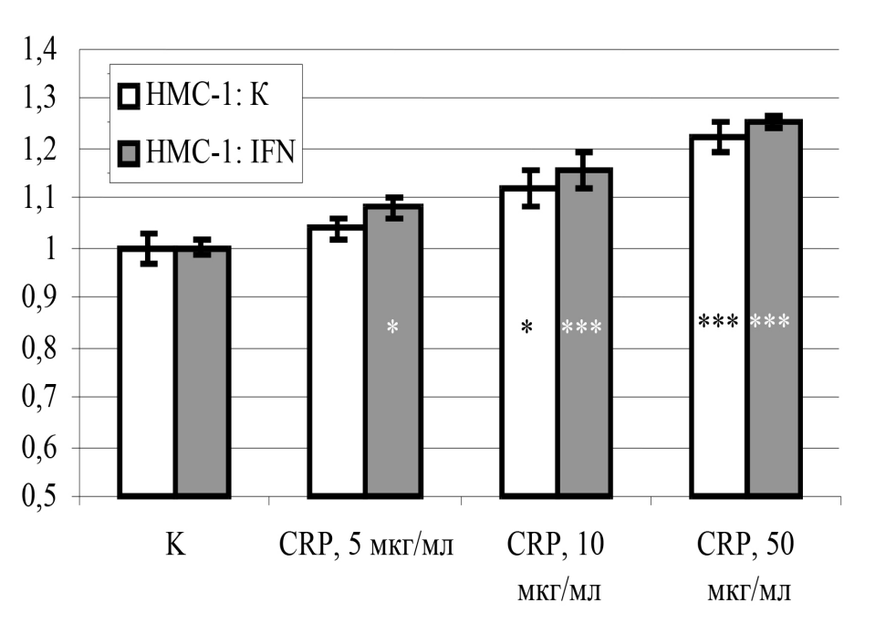
Fig. 9. Adhesion of HMC-1 mast cells to fibroblasts treated with CRP. Role of HMC-1 cell activation by IFNγ.
Note: the x-axis shows CRP concentration; K is the control (without CRP treatment). The y-axis shows the stimulation index. White bars — intact MCs; gray bars — MCs stimulated with IFNγ for 24 hours. Differences from control are significant: * — p < 0.05; *** — p < 0.001.
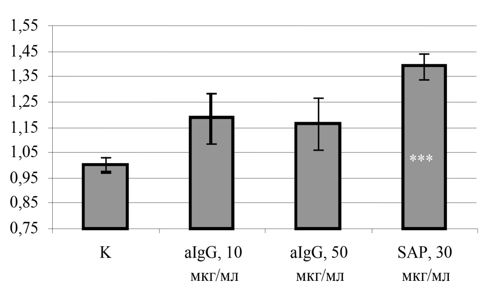
Fig. 10. Adhesion of HMC-1 mast cells to fibroblasts treated with aIgG or SAP
Note: the ordinate shows the stimulation index. *** — differences from control are significant, p < 0.001.
Based on the increased FcγRI expression observed on HMC-1 MCs after IFNγ treatment, the effect of CRP on the adhesion of IFNγ-activated cells was investigated. CRP stimulation at 5, 10, and 50 μg/ml enhanced adhesion of IFNγ-treated MCs by 8 %, 15 %, and 25 %, respectively (p < 0.05) (Fig. 9). IFNγ alone had no effect on MC adhesion to Fb, even in the presence of CRP.
The results showed that even at a CRP dose of 5 μg/ml, there was a significant 8 % increase in the fluorescence of adherent, interferon-activated MCs (p < 0.05). At a CRP concentration of 10 μg/ml, the increase in adhesion reached 15 %, and at 50 μg/ml, 25 %. The effects of CRP at concentrations of 5, 10, and 50 μg/ml differed significantly from the control (p < 0.05) (Fig. 9).
As shown in Figure 9, CRP enhances the adhesion of IFNγ-pretreated MCs to Fb. At the same time, IFNγ itself has almost no effect on MC adhesiveness to Fb in the presence of CRP (Fig. 9). The only robust finding is that the presence of CRP leads to higher adhesion. Just as IFNγ does not affect CRP binding to MCs, it does not affect MC adhesion to CRP-treated fibroblasts. The only difference is that, at the lowest concentration, the difference from control is significant for IFNγ-treated cells.
Thus, the data obtained indicate that C-reactive protein affects not only cells of the immune system but also connective tissue fibroblasts, increasing their adhesiveness for mast cells. The mechanism underlying the effect of CRP on fibroblast adhesiveness is most likely different from that in mast cells, since fibroblasts do not express the Fc receptors through which the pentraxin usually acts on cells. This mechanism requires separate investigation and is discussed below.
In contrast to intact MCs, IFNγ-stimulated MCs interact only slightly more strongly with CRP-stimulated Fb.
Stimulation of the Fb monolayer with aIgG at concentrations of 10 and 50 μg/ ml also produced a significant increase in the adhesion of IFNγ-stimulated HMC- 1 cells, by 20 % and 10 %, respectively. The differences between the different aIgG concentrations were significant (p < 0.01) (Fig. 11).
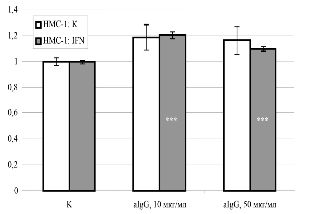
Fig. 11. Effect of aIgG on the adhesive properties of fibroblasts
Note: aIgG was added to fibroblasts at the indicated concentrations and incubated for 24 hours. Intact (white bars) or 24-hour IFNγ-stimulated (gray bars) HMC-1 cells were then added to the fibroblasts. The ordinate shows the stimulation index; *** — differences from control are significant, p < 0.001. Control (C) — adhesion of unstimulated MCs (white bar) or IFNγ-stimulated MCs (gray bar) to intact fibroblasts.
A somewhat different pattern was observed when, before being added to a CRP-stimulated Fb monolayer, the Fcγ receptors of HMC-1 MCs were “blocked” with IgG Fc fragments; for this purpose, HMC-1 cells were incubated for 20 min on ice in PBS containing either Fc fragments or whole IgG molecules at a concentration of 25 μg/ml. Blocking MC Fcγ receptors with IgG Fc fragments (25 μg/ml, 20 min, 4 °C) reduced their adhesion to CRP-stimulated Fb to the level of spontaneous attachment to intact cells, whereas adhesion to TNF-activated Fb remained unchanged (Fig. 12, a). Similarly, pretreatment of IFNγ-stimulated MCs with Fc fragments completely abolished the CRP-dependent enhancement of adhesion (Fig. 12, b).
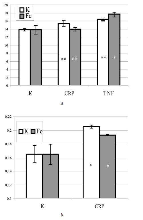
Fig. 12. Adhesion of HMC-1 mast cells to fibroblasts activated with CRP. Role of Fcγ receptors on HMC-1 cells
Note: fibroblasts were activated with CRP at 50 μg/ml, after which either intact MCs (white bars) or MCs pre-incubated with IgG Fc fragments (gray bars) were added: a — MCs not stimulated with IFNγ; b — HMC-1 MCs activated with IFNγ for 24 hours. The ordinate shows RFU. Significance of differences from control: * — p < 0.05; *** — p < 0.001; significance of differences between intact MCs and MCs treated with IgG Fc fragments: # — p < 0.05, ## — p < 0.01.
The same blockade of mast cell FcγR had no effect on their adhesion to TNF-stimulated Fb.
6.3. Effect of C-reactive protein on the adhesion of HMC-1 mast cells
to fibroblasts in its presence
This part of the study examined the effect of C-reactive protein and aggregated IgG on the adhesion of HMC-1 mast cells to fibroblasts when present during co-culture. Unlike the standard protocol, the cell populations under study were not pre-stimulated with C-reactive protein; instead, it was added directly to the mixture of interacting cells for the entire duration of co-culture.
The experimental data showed that adding CRP at 50 μg/ml to the co-culture medium enhanced mast cell adhesion to fibroblasts. A clear relationship was observed between adhesion level and incubation time: the number of attached cells increased over the 20—50 min interval. The dynamics of the process showed a peak of activity within the first 30 min. At 20 min after the start of incubation, adhesion-related fluorescence increased by 25 % relative to control samples (without CRP). The maximum effect was recorded at 30 min, when the number of attached cells reached 186 % of control, corresponding to an 86 % increase. However, with further prolongation of co-culture, the differences between experimental and control groups decreased: 22 % at 40 min and only 11 % at 50 min (Fig. 13).
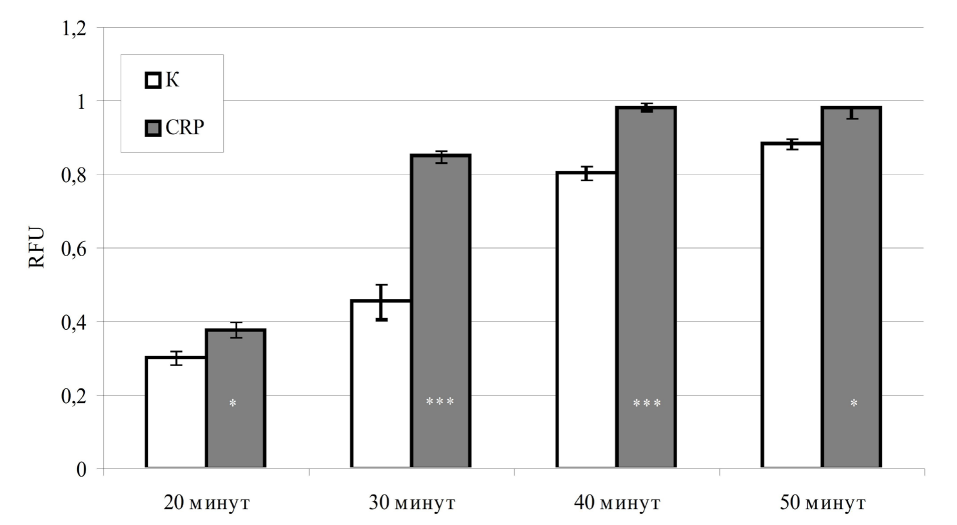
Fig. 13. Adhesion of HMC-1 mast cells to Fb during co-culture of a cell mixture in the presence of C-reactive protein. Time dependence
Note: white bars — spontaneous adhesion of mast cells to Fb; gray bars — adhesion in the presence of CRP, 50 μg/ml. Significance of differences from control: * — p < 0.05, *** — p < 0.001.
6.4. Effect of C-reactive protein on the adhesiveness of EA.hy926 endothelial cells for HMC-1 mast cells
Experiments demonstrated that activation of EA.hy926 endothelial cells with C-reactive protein (CRP) enhanced the adhesion of HMC-1 mast cells. The effect of CRP was dose-dependent: at a concentration of 10 μg/ml, adhesive activity increased by 50 %, and at 50 μg/ ml it reached 80 % relative to control (unstimulated endothelial cells). Statistical analysis confirmed the significance of the differences between the effects of CRP at these concentrations (p < 0.05) (Fig. 14).
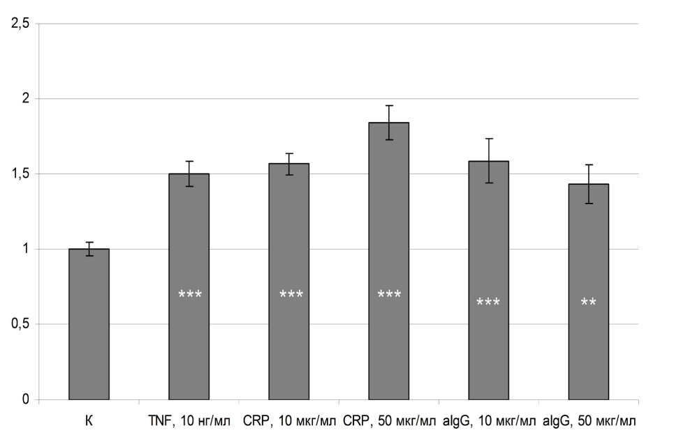
Fig. 14. Effect of C-reactive protein and aggregated IgG on the adhesive properties of EA.hy926 endothelium for HMC-1 mast cells
Note: the ordinate shows the stimulation index. Differences from control are significant: ** — p < 0.01, *** — p < 0.001.
A similar enhancement of adhesion was recorded upon stimulation of the endothelium with aggregated IgG (aIgG). At an aIgG concentration of 10 μg/ml, the fluorescence intensity reflecting the number of attached mast cells increased by 60 %, and at 50 μg/ml by 43 %. Both values differed significantly from the negative control (adhesion to intact endothelium, p < 0.05); however, the difference between the groups treated with different aIgG doses did not reach statistical significance (p > 0.05) (Fig. 14).
In addition, under the experimental conditions used, adhesion of HMC-1 mast cells to endothelium activated with the pro-inflammatory cytokine TNF-α (10 ng/ml) increased 1.5-fold compared with control (Fig. 14).
7. Effect of C-reactive protein on activation of the transcription factor NF-κB in HMC-1 mast cells
HMC-1 cells transfected with the pNF-κBluc plasmid responded to CRP stimulation with increased luciferase synthesis: luminescence in CRP-stimulated cells increased by 40 % compared to control cells (Fig. 15).
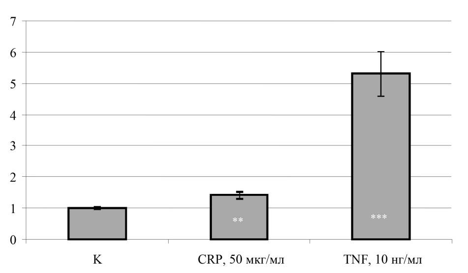
Fig. 15. Effect of human C-reactive protein on expression of the luciferase reporter gene driven by the NF-κB promoter in HMC-1 cells transfected with the pNF-κBluc construct
Note: the ordinate shows luciferase luminescence intensity in relative luminescence units (RLU); ** — differences from control are significant, p < 0.01; *** — differences from control are significant, p < 0.001.
Discussion
The results of this study confirm that HMC-1 mast cells express the low-affinity Fcγ receptor CD32 (FcγRII). According to the literature, intact human MCs derived from CD34+ blood cells also express CD32 but contain minimal amounts of CD64 (FcγRI) and CD16 (FcγRIII) mRNA in the cytoplasm. Stimulation with interferon-γ (IFNγ) induces expression of the high-affinity FcγRI (CD64), while expression of the low-affinity receptors remains unchanged [11]. Our data are consistent with this: intact HMC-1 MCs show baseline CD64 expression, which is enhanced by IFNγ. Maximal FcγRI expression was recorded 24 hours after stimulation, after which its level decreased.
It should be noted that several studies report that CD32 is the only Fcγ receptor on HMC-1 MCs [12]. In our experiments, we did not have access to antibodies capable of distinguishing between the FcγRII subtypes (CD32a, CD32b, and CD32c), and therefore cannot state with certainty which CD32 subtype is expressed on HMC-1 cells. The inhibitory receptor CD32b may be absent from human MCs, in contrast to blood basophils. In mouse MCs, by contrast, CD32b is expressed, and its activation generates an inhibitory signal that suppresses activation induced by aggregation of FcεRI (the high-affinity IgE receptor) [13]. In our experiments, both aIgG and CRP predominantly activated MCs and, in any case, did not suppress any MC function; we therefore conclude that aIgG and CRP interact with an activating rather than an inhibitory Fcγ receptor.
The experiments revealed dose-dependent binding of CRP to the MC surface. Interestingly, binding was enhanced in the absence of Ca2+ and Mg2+. CRP, being a calcium-dependent protein, undergoes a conformational change upon saturation with Ca2+ ions, which regulates its interaction with ligands. For example, binding to phosphorylcholine requires Ca2+, whereas interaction with polycations / polyanions is suppressed [10]. This suggests that CRP binds to Fcγ receptors through sites unrelated to the phosphorylcholine-specific sites.
The literature indicates that calcium ions regulate the interaction of the CRP molecule with different ligands, switching the pentraxin’s specificity from one ligand to another [10]. For example, interaction of CRP with phosphorylcholine (PC)-containing ligands (such as pneumococcal C-polysaccharide) requires saturation of the CRP molecule with calcium ions, which opens the PC-specific binding sites and simultaneously exposes the C1q-binding site. Consequently, a CRP molecule bound to a PC-containing ligand recruits C1q and activates complement via the classical pathway. In contrast, pentraxin binding to ligands of another type, such as polycations and polyanions (e. g., histones, polyribonucleotides), is inhibited by calcium ions: when the CRP molecule is saturated with calcium, the charged peptide regions of CRP responsible for contact with polycations and polyanions are no longer exposed on the surface of the molecule, so binding to these ligands is blocked. Interaction of CRP with polycations/polyanions is also not accompanied by complement activation. Given the regulatory role of calcium ions in exposing the PC-binding and charged sites of CRP, it can be concluded that CRP binds to Fcγ receptors through sites unrelated to the phosphorylcholine-specific sites, and that this binding most likely occurs via the opposite face of the CRP molecule, where clusters of charged amino acid residues are located [10].
The resolution of the method allowed detection of biotin-labeled CRP binding to MCs starting from 12.5 μg/ml (flow cytometry, APC-conjugated streptavidin). At CRP concentrations above 100 μg/ml, no further increase in binding to mast cells occurred, most likely reflecting saturation of the receptors capable of interacting with CRP. In the presence of either unlabeled CRP or IgG, binding of C-reactive protein to cells decreased, indicating the specificity of binding and a shared receptor for CRP and aIgG on mast cells (see Fig. 1, c, d ).
Although IFNγ increased FcγRI expression on MCs, it had no effect on CRP binding to MCs. This may be explained by the fact that FcγRII is present on cells in much greater numbers (see Fig. 2), so that the increase in FcγRI expression produces only a negligible change in CRP binding when measured by flow cytometry.
It has been shown that stimulation of FcγRII on human mast cells does not induce histamine release. In addition, stimulation of HMC-1 with aIgG has been shown not to induce TNF release [12].
Thus, under normal conditions, FcγRII is the only receptor for immunoglobulin G on human mast cells, meaning that IgG stimulation of MCs either fails to induce degranulation at all or induces it only to a minor degree. It is known that, following IFNγ stimulation, MCs acquire the ability to respond to IgG [14]. Our data are consistent with this. Intact HMC-1 cells did not respond to CRP added to the medium with significant histamine release. However, when cells had been activated with IFNγ for 24 hours, histamine release was observed in response to CRP stimulation. According to the literature, the aIgG concentration at which MC degranulation is maximal is 1 μg/ml [14]. In our experiments, the greatest histamine release was observed with CRP stimulation at a concentration of 3 μg/ml, i. e., at a CRP concentration of the same order of magnitude as that reported for aIgG-induced degranulation.
MCs are capable of degranulation not only upon FcεRI aggregation but also upon aggregation of CD64 (FcγRI). FcγRI-dependent degranulation can be induced not only by antibodies to the receptor but also by the addition of aIgG, or by prior “sensitization” with human IgG1 followed by the addition of murine anti-human IgG1 antibodies [14]. Notably, the IgG2 and IgG3 subclasses, together with the corresponding anti-IgG antibodies, were unable to induce MC degranulation. Particularly interesting is the finding that IgG3 was unable to induce degranulation of IFNγ-stimulated MCs, even though this subclass has an affinity for FcγRI comparable to that of IgG1. Histamine release from MCs induced by IgG4/anti-IgG4 did occur, but was an order of magnitude weaker than that induced by IgG/ IgG1 [14].
The literature describes MC degranulation in affected tissue areas in the presence of immune complex deposition. Activated MCs have been found, for example, in the inflamed synovium of patients with rheumatoid arthritis and scleroderma, and in deposits in IgG-associated vasculitis [14]. Elevated tissue IFNγ levels are known to be characteristic of a number of bacterial and viral infections [15]. This clarifies the mechanism by which MCs become involved in the pathogenesis of such diseases. For example, elevated levels of IFNγ and MCs are observed at lesion sites in psoriasis [16]. In addition, FcγRI has been shown to be expressed by these cells [17]. It is possible that MCs are thereby involved in tissue damage in conditions where a pathological role of MCs, together with elevated levels of immune complexes and CRP, has been demonstrated — for example, in psoriasis [16] and rheumatoid arthritis [18].
Thus, an antigen-dependent MC response is possible not only in the context of a Th2-associated immune response, in which IL-4 and IL-13 drive IgE synthesis, but also in a Th1 response, in which secreted IFNγ increases CD64 expression on cells and promotes IgG synthesis.
As our experiments showed, CRP and aIgG at concentrations of 10 and 50 μg/ml stimulated the adhesive properties of mast cells. The literature indicates that C-reactive protein characteristically exhibits pro-inflammatory activity [10]. Furthermore, aggregation of Fcγ receptors on the surface of murine mast cells has been shown to enhance their adhesion to fibronectin [19].
MCs are known to be consistently present in fibrotic tissue and to be associated with the development of fibrosis, although the mechanisms linking MCs to fibrosis remain insufficiently understood. MCs have been shown to stimulate profibrotic activity in fibroblasts by forming contacts with fibroblasts, promoting their proliferation, and inducing contraction of the collagen network [20]. MCs also influence fibroblast expression of receptors for histamine, serotonin, TNF, and tryptase, as well as fibroblast release of the chemokines CCL8, CCL13, CXCL4, and CXCL6 [20].
As our experiments showed, stimulation of fibroblasts with C-reactive protein nevertheless results in some enhancement of mast cell adhesion to them (see Fig. 9), despite the absence of CRP receptors (i. e., Fcγ receptors) on fibroblasts. Fibroblasts growing in culture are known to produce matrix proteins, creating a protein microenvironment around themselves. CRP has binding sites for various connective tissue matrix proteins, in particular fibronectin and laminin [9; 10]. The Fcγ receptor-binding sites, in turn, are located on the opposite face of the CRP molecule [10]. Based on this, we hypothesized that pre-incubation of fibroblasts with CRP may promote their adhesion to mast cells indirectly, through CRP binding to components of the extracellular matrix (fibronectin and other proteins), rather than through direct binding to and activation of fibroblasts. When mast cells are subsequently added, they in turn bind, via their FcγR, to the matrix-associated CRP, thereby enhancing adhesion. This hypothesis is consistent with the fact that the CRP sites responsible for contact with the matrix proteins fibronectin and laminin are the PC-specific sites located on one face of the flat, discoidal CRP molecule, while the Fcγ receptor-binding sites are located on the other [10].
This is confirmed by the results of the following experiment. Before being added to Fb that had been incubated with CRP at 50 μg/ml (or with TNF at 10 ng/ml) for 24 hours, MCs were treated with IgG Fc fragments to “block” their Fcγ R. Adhesion of Fc fragment-treated mast cells to CRP-activated fibroblasts was significantly lower than that of MCs with unblocked Fcγ receptors, and was at the level of adhesion of intact MCs to untreated fibroblasts, whereas blocking MC Fcγ receptors had no effect on the degree of MC attachment to TNF-activated Fb (see Fig. 12).
If CRP acts as an anchor in MC adhesion to Fb, it could be predicted that co-incubating Fb and MCs in the presence of CRP would increase the intensity of MC adhesion to Fb. Indeed, co-incubation of MCs and Fb in the presence of CRP resulted in a significant increase in the degree of adhesion (see Fig. 13).
Thus, the hypothesis that C-reactive protein does not activate fibroblasts but rather serves as an anchor for MCs within the connective tissue matrix was confirmed. It has been described in the literature that the membrane-bound form of CRP expressed by human monocytes can function as a cell adhesion molecule, mediating the interaction between monocytes and fibronectin [21]. It is also known that CRP binding to fibronectin interferes with Fb binding to fibronectin, and that this inhibition is Ca2+-dependent [10]. The Ca2+ dependence of this inhibition indicates that CRP binds to fibronectin via the epitope responsible for its interaction with PC (inhibition of CRP—fibronectin binding by phosphorylcholine was demonstrated in the same study); the Fcγ receptor-binding site is located on the opposite face of the molecule, but since fibroblasts lack Fcγ receptors, Fb adhesion to fibronectin is inhibited. At the same time, according to our data, cells expressing Fcγ receptors on their surface show, conversely, more active adhesion to extracellular matrix proteins in the presence of CRP (Fig. 16).
It has been reported in the literature that CRP induces NF-κB activation in cultured human glomerular mesangial cells. At concentrations of 12.5—100 μg/ ml, CRP dose-dependently increased the DNA-binding capacity of nuclear NF-κB, with a maximum at 3 hours after incubation of cells with CRP in vitro. Incubation of mesangial cells with CRP also enhanced NF-κB-dependent mRNA expression of monocyte chemoattractant protein-1 (MCP-1). CRP also increased intracellular calcium concentration and the generation of reactive oxygen species [22].
Studies on the role of CRP in atherogenesis and the development of cardiovascular complications have shown that CRP acts on vascular wall cells as a pro-inflammatory inducer, driving endothelial cells to acquire a so-called inflammatory phenotype and pro-atherogenic properties. In particular, its pro-inflammatory effect on endothelial cells has been shown to manifest as increased expression of the adhesion molecules VCAM-1 and CD40, which mediate leukocyte recruitment and attachment to the vessel wall. CRP stimulated VCAM-1 mRNA expression, VCAM-1 protein synthesis and expression, and CD40 expression, and activated the VCAM-1 gene promoter; these effects of CRP involved protein kinase C, the mitogen-activated protein kinase p38MAPK, tyrosine kinase, and the transcription factor NF-κB. Inhibitors of these kinases and NF-κB inhibitors (parthenolide, pyrrolidine dithiocarbamate) abolished the induction of VCAM-1 by C-reactive protein [23].
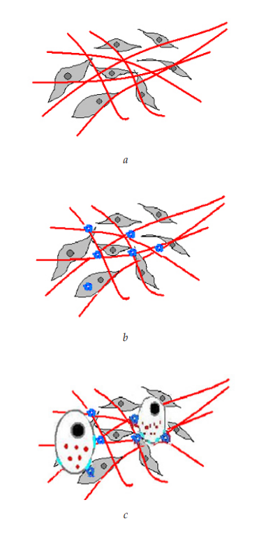
Fig. 16. CRP stimulation of cell adhesion to the extracellular matrix:
a — Fibroblasts and the extracellular matrix they synthesize;
b — CRP bound to extracellular matrix proteins (e. g., fibronectin and laminin)
via the face bearing the Ca2+-dependent ligand-binding sites;
c — Cells interacting with CRP via FcγR anchor themselves in tissue
It has been shown that the CRP-induced, NF-κB-mediated increase in VCAM-1 expression by aortic endothelial cells in vitro involves type II Fcγ receptors. This was confirmed by inhibiting CD32 mRNA using several small interfering RNAs (siRNAs) targeting a 21-nucleotide region of CD32 mRNA. Cells incubated with the siRNAs did not express CD32 and did not show increased VCAM-1 expression in response to C-reactive protein stimulation [23].
The NF-κB-dependent pathway is also activated by other Fc receptor ligands, in particular by their classical ligand, immunoglobulin G. Thus, in human monocyte-derived dendritic cells, activation of type II Fcγ receptors (FcγRII, CD32) by aggregated (immobilized) IgG has been shown to induce differentiation and cytokine synthesis (IL-10), as well as the acquisition of the ability to induce proliferation of allogeneic T cells. These dendritic cell responses to FcγRII cross-linking involve the NF-κB-dependent pathway, with translocation of the NF-κB proteins p65 and c-Rel from the cytoplasm to the nucleus. The dendritic cell response to IgG was abolished in the presence of blocking anti-CD32 antibodies [24].
It has been shown that stimulation with C-reactive protein enhances the adhesive properties of mast cells and endothelial cells (see Fig. 7, 14; Table 3). As shown by our data (see Fig. 1, d) and consistent with the literature, C-reactive protein and aIgG share the same receptors — type I and II FcγR [25]. Likewise, our experimental results demonstrated a stimulatory effect of aIgG on cell adhesion. It can therefore be concluded that FcγRII activation triggers a signaling cascade resulting in the activation of genes responsible for cell adhesion to one another or to other substrates, such as connective tissue matrix proteins. A similar effect of CRP has been demonstrated, in particular, in endothelial cells isolated from the aorta [25]. In addition, the authors of this study showed that CD32 gene silencing by siRNA reduced the adhesive properties of endothelial cells relative to intact CRP-treated cells. The same effect was achieved by adding blocking antibodies against CD32 or CD64 to the endothelial cell culture medium.
Table 3
Effect of C-reactive protein and aggregated IgG on the adhesion
of the studied cell lines (percentage increase relative to control)
Stimulated cells | Adhesion partner (untreated) | CRP | aIgG |
HMC-1 | Fibroblasts | 54 % | 60 % |
EA.hy926 | HMC-1 | 80 % | 43 % |
Note: CRP and aIgG were used at a concentration of 50 μg/ml. Control: adhesion level for the corresponding pair of untreated cells.
Thus, our experiments demonstrated that the main receptors for CRP on EA.hy926 endothelial cells and HMC-1 mast cells are the low-affinity IgG receptors, FcγRII, as previously reported in the literature for endothelial cells isolated from the aorta or umbilical vein.
Moreover, by acting through FcγR on MCs, CRP triggers a chain of intracellular processes that alter the adhesive properties of these cells independently of increased expression of the adhesion molecules CD54 and CD11b. Consequently, the mechanism underlying CRP- and aIgG-induced enhancement of MC adhesiveness still requires further investigation.
The high-affinity receptor FcγRI also serves as a CRP receptor on immunocompetent cells but, unlike FcγRII, which is expressed by virtually all cells of hematopoietic origin and by endothelium, it is normally restricted to monocytes and macrophages; mast cells begin to express it only after IFNγ stimulation. IFNγ stimulation of HMC-1 MCs induces CD64 expression, and CRP-mediated activation of this receptor leads to MC degranulation.
Reference
1. Crivellato E., Ribatti D. The mast cell: an evolutionary perspective. Biol. Rev. Camb. Philos. Soc. 2010, 85(2), 347—360, DOI: 10.1111/j.1469-185x.2009.00105.x.
2. Rivellese F., Rossi F. W., Galdiero M. R., Pitzalis C., de Paulis A. Mast Cells in Early Rheumatoid Arthritis. Int. J. Mol. Sci. 2019, 20(8), 2040, DOI: 10.3390/ijms20082040.
3. Ribatti D., Tamma R., Annese T. Mast cells and angiogenesis in multiple sclerosis. Inflamm. Res. 2020, 69(11), 1103—1110, DOI: 10.1007/s00011-020-01394-2.
4. Conti P., Caraffa A., Mastrangelo F., Tettamanti L., Ronconi G., Frydas I., et al. Critical role of inflammatory mast cell in fibrosis: Potential therapeutic effect of IL-37. Cell Prolif. 2018, 51(5), e12475, DOI: 10.1111/cpr.12475.
5. Weber A., Knop J., Maurer M. Pattern analysis of human cutaneous mast cell populations by total body surface mapping. British Journal of Dermatology. 2003, 148(2), 224—228, DOI: 10.1046/j.1365-2133.2003.05090.x.
6. Huttenlocher A., Sandborg R. R., Horwitz A. F. Adhesion in cell migration. Curr. Opin. Cell Biol. 1995, 7(5), 697—706, DOI: 10.1016/0955-0674(95)80112-x.
7. Dileepan K. N., Raveendran V. V., Sharma R., Abraham H., Barua R., Singh V., et al. Mast cell-mediated immune regulation in health and disease. Front. Med. 2023, 10, 1213320, DOI: 10.3389/fmed.2023.1213320.
8. Ma Y. J., Parente R., Zhong H., Sun Y., Garlanda C., Doni A. Complement-pentraxins synergy: Navigating the immune battlefield and beyond. Biomed. Pharmacother. 2023, 169, 115878, DOI: 10.1016/j.biopha.2023.115878.
9. Singh B., Goyal A., Patel B. C. C-Reactive Protein: Clinical Relevance and Interpretation. In: StatPearls [Internet]. StatPearls Publishing; 2025.
10. Nazarov P. G. Acute-phase reactants of inflammation [Reaktanty ostroy fazy vospaleniya]. Nauka, 2001, ISBN 5-02-026122-Х. (In Russ.)
11. Okayama Y., Kirshenbaum A. S., Metcalfe D. D. Expression of a functional high-affinity IgG receptor, Fc gamma RI, on human mast cells: up-regulation by IFN-gamma. The Journal of Immunology. 2000, 164(8), 4332—4339, DOI: 10.4049/jimmunol.164.8.4332.
12. Wedi B., Lewrick H., Butterfield J. H., Kapp A. Human HMC-1 mast cells exclusively express the Fc gamma RII subtype of IgG receptor. Arch. Dermatol. Res. 1996, 289(1), 21—27, DOI: 10.1007/s004030050147.
13. Kanagaratham C., Derakhshan T., El Ansari Y. S., Furiness K. N., Hollers E., Keldsen M., et al. IgG:FcγRIIb signals block effector programs of IgE:FcεRI-activated mast cells but spare survival pathways. J. Allergy Clin. Immunol. 2023, 152(2), 453—468, DOI: 10.1016/j.jaci.2023.03.027.
14. Woolhiser M. R., Okayama Y., Gilfillan A. M., Metcalfe D. D. IgG-dependent activation of human mast cells following up-regulation of Fc gamma RI by IFN-gamma. Eur. J. Immunol. 2001, 31(11), 3298—3307, DOI: 10.1002/1521-4141(200111)31:11 < 3298::aid-immu3298 > 3.0.co;2-u.
15. Jouanguy E., Doffinger R., Dupuis S., Pallier A., Altare F., Casanova J. L. IL-12 and IFN-gamma in host defense against mycobacteria and salmonella in mice and men. Curr. Opin. Immunol. 1999, 11(3), 346—351, DOI: 10.1016/s0952-7915(99)80055-7.
16. Sieminska I., Pieniawska M., Grzywa T. M. The Immunology of Psoriasis — Current Concepts in Pathogenesis. Clin. Rev. Allergy Immunol. 2024, 66(2), 164—191, DOI: 10.1007/s12016-024-08991-7.
17. Tkaczyk C., Okayama Y., Woolhiser M. R., Hagaman D. D., Gilfillan A. M., Metcalfe D. D. Activation of human mast cells through the high affinity IgG receptor. Mol. Immunol. 2002, 38(16—18), 1289—1293, DOI: 10.1016/s0161-5890(02)00077-9.
18. Pope J. E., Choy E. H. C-reactive protein and implications in rheumatoid arthritis and associated comorbidities. Semin. Arthritis Rheum. 2021, 51(1), 219—229, DOI: 10.1016/j.semarthrit.2020.11.005.
19. Pastwińska J., Walczak-Drzewiecka A., Łukasiak M., Ratajewski M., Dastych J. Hypoxia regulates human mast cell adhesion to fibronectin via the PI3K/AKT signaling pathway. Cell Adh. Migr. 2020, 14(1), 106—117, DOI: 10.1080/19336918.2020.1764690.
20. Foley T. T., Saggers G. C., Moyer K. E., Ehrlich H. P. Rat mast cells enhance fibroblast proliferation and fibroblast-populated collagen lattice contraction through gap junctional intercellular communications. Plast. Reconstr. Surg. 2011, 127(4), 1478—1486, DOI: 10.1097/PRS.0b013e318208d0bb.
21. Kolb-Bachofen V., Puchta-Teudt N., Egenhofer C. Expression of membrane-associated C-reactive protein by human monocytes: indications for a selectin-like activity participating in adhesion. Glycoconj. J. 1995, 12(2), 122—127, DOI: 10.1007/BF00731355.
22. Chang J. W., Kim C. S., Kim S. B., Park S. K., Park J. S., Lee S. K. C-reactive protein induces NF-kB activation through intracellular calcium and ROS in human mesangial cells. Nephron Exp. Nephrol. 2005, 101(4), e165—172, DOI: 10.1159/000087940.
23. Liang Y.-J., Shyu K.-G., Wang B.-W., Lai L.-P. C-reactive protein activates the nuclear factor-kB pathway and induces vascular cell adhesion molecule-1 expression through CD32 in human umbilical vein endothelial cells and aortic endothelial cells. J. Mol. Cell Cardiol. 2006, 40(3), 412—420, DOI: 10.1016/j.yjmcc.2005.12.008.
24. Banki Z., Kacani L., Mullauer B., Wilflingseder D., Obermoser G., Niederegger H., et al. Cross-linking of CD32 induces maturation of human monocyte-derived dendritic cells via NF-kB signaling pathway. J. Immunol. 2003, 170(8), 3963—3970, DOI: 10.4049/jimmunol.170.8.3963.
25. Bharadwaj D., Stein M. P., Volzer M., Mold C., Clos T. W. D. The major receptor for C-reactive protein on leukocytes is Fcγ receptor II. J. Exp. Med. 1999, 190(4), 585—590, DOI: 10.1084/jem.190.4.585.